


GeneMedi's protocol / procedure for the diagnostics application-CLIA
1 What is Chemiluminescence Immunoassay
Chemiluminescence (CL) is defined as the emission of electromagnetic radiation caused by a chemical reaction to produce light.
Chemiluminescence immunoassay (CLIA) is an assay that combine chemiluminescence technique with immunochemical reactions. Similar with other labeled immunoassays (RIA, FIA, ELISA), CLIA utilize chemical probes which could generate light emission through chemical reaction to label the antibody.
Chemiluminescent immunoassay (CLIA) is an immunoassay technique where the label, i.e. the true "indicator" of the analytic reaction, is a luminescent molecule.
In general, luminescence is the emission of visible or near-visible ( λ = 300–800 nm) radiation which is generated when an electron transitions from an excited state to ground state. The resultant potential energy in the atom gets released in the form of light.
In spectrophotometry, luminescence has an advantage over absorbance in that the former is an absolute measure whereas the latter is relative.
We make reference to chemiluminescence, because the type of luminescence applied to immunoassay techniques generally identifies exergonic chemical reactions as the most suitable energy source for producing the electronic excited state.
Chemiluminescent methods can be direct—using luminophore markers—or indirect—using enzyme markers. Either method may be competitive or non-competitive.
In direct chemiluminescent methods, the luminophore markers used are acridinium and ruthenium esters, while the enzymatic markers used in indirect methods are alkaline phosphatase with adamantyl 1, 2-dioxetane aryl phosphate (AMPPD) substrate and horseradish peroxidase with luminol or its derivatives as substrate.
Synthesizing molecules such as AMPPD and isoluminol base molecule derivatives are more stable compared to other luminescent markers and result in light emission with a characteristically elevated quantum yield. Activation of these substrates requires chemical or enzymatic reactions associated with the immunological reaction.
For example, the use of luminol and derivatives of isoluminol as chemiluminescent labels depends on the coupling of the immunoassay with enzymatic reactions catalyzed by peroxidase. The addition of an enhancer (e.g. ferrocyanide, metallic ions) further boosts the electronic activation, ultimately leading to extremely elevated analytic sensitivity (mol per litre), certainly superior to those attainable by other immunoassay methods such as RIA, immunoenzymatic (ELISA) and fluoroimmunoenzymatic (FEIA) methods, etc.
2 Four types of Chemiluminescence Immunoassay
2.1 Chemiluminescence immunoassay
Chemiluminescence immunoassay (CLIA) is a type of immunoassay method that directly label antigens or antibodies with chemiluminescence agents. Acridine esters are ideal luminescent substrates, which can be oxidized by hydrogen peroxide to emit light in an alkaline environment.Chemiluminescence (CL) is defined as the emission of electromagnetic radiation caused by a chemical reaction to produce light.
2.2 Chemiluminescence enzyme immunoassay
Chemiluminescence Enzyme Immunoassay (CLEIA) is an enzyme-labeled immunoassay that uses chemiluminescence agents as substrates for enzyme reactions. After two-stage amplification of enzyme and luminescence, it has high sensitivity. Peroxidase is used as a labeling enzyme, luminol is used as a luminescent substrate, and a luminescence enhancer is added to improve sensitivity and luminescence stability. The applied labeling enzyme can also be alkaline phosphatase, the luminescent substrate is dioxetane phosphate, and the solid phase carrier is magnetic particles.
2.3 Microparticle chemiluminescence immunoassay
The immunoassay technology has two methods: one is the determination of small molecule antigenic substances by the competition method; the other is the measurement of antigenic substances of large molecules by the double antibody sandwich method.
The solid-phase magnetic powder particles used in this instrument are extremely small, with a diameter of only 1.0 μm. This greatly increases the coating surface area, increases the amount of antigen or antibody adsorption, speeds up the reaction, and makes cleaning and separation easier.
The basic reaction process:
(1) Competitive reaction: Use an excess of antibody coated with magnetic particles, and add the antigen to be tested and the quantitative labeled acridinium ester antigen to the reaction cup for incubation. There are two types of binding of the immune response. One is to bind the labeled antigen and antibody to form a complex, and the other is to determine the binding of antigen and antibody.
(2) Double-antibody sandwich method: The labeled antibody and the test antigen are combined with the coating antibody to form a reaction form, that is, a complex of the coating antibody-determining antigen-luminescent antibody.
2.4 Electro chemiluminescence immunoassay
Electrochemiluminescence immunoassay (ECLI) is a specific luminescence reaction initiated by electrochemistry on the electrode surface, including electrochemistry and chemiluminescence. The label used in the analysis is the electrochemiluminescence substrate ruthenium terpyridine or its derivative N-hydroxysuccinamide (NHS) ester, which can be combined with antibodies or antigen molecules of different chemical structures through chemical reactions to produce labeled antibodies or antigens.
3 Advantages and disadvantages of chemiluminescence immunoassay
Chemiluminescence immunoassay has unique characteristics of high sensitivity, high specificity, speed, accuracy, specificity, and automation.
It plays an important role in clinical testing, drug analysis, environmental monitoring and other fields. Especially the determination of various hormones, tumor markers, drug concentration and other trace biologically active substances.
In practical analysis, it is necessary to detect complex samples with large amounts and low abundance, so researchers have followed the trend to develop rapid, high-throughput and sensitive CLIA.
The time for measuring the optical signal of each sample is no more than a few seconds, and it can be completed within 9-60 minutes from sample addition to analysis result.
Additionally, CLIA has a wide dynamic range, which means that it can accurately measure a broad range of concentrations in a single assay. Another advantage of CLIA is that it is a homogeneous assay, meaning that it does not require any separation or washing steps, making it a fast and efficient assay.
However, CLIA also has some disadvantages. One limitation of CLIA is that it requires specialized equipment and reagents, which can be costly and may not be available in all laboratories. Another limitation to consider is the lot-to-lot variability, an issue for all assays.
Lot-to-lot differences in the reagents can be due to variability of raw materials used in the formulation of the reagents. This can impact the quality of the assay and must be carefully analyzed. On the other hand, It is important to check lot-to-lot differences in the calibrations and controls because response of certain reagents may change.
Although the development of a chemiluminescent immunoassay requires the optimization of several key parameters, with the help of solid expertise of researchers in the field, improved development of chemiluminescent immunoassays can be developed.
Additionally, CLIA may be subject to interference from matrix effects, such as high levels of lipids or proteins in a sample, which can lead to false-positive or false-negative results.
The other disadvantages are:
- the light-emitting process is short;
- High background;
- High instrument failure rate;
- The detection accuracy is not high
4 Test Procedure
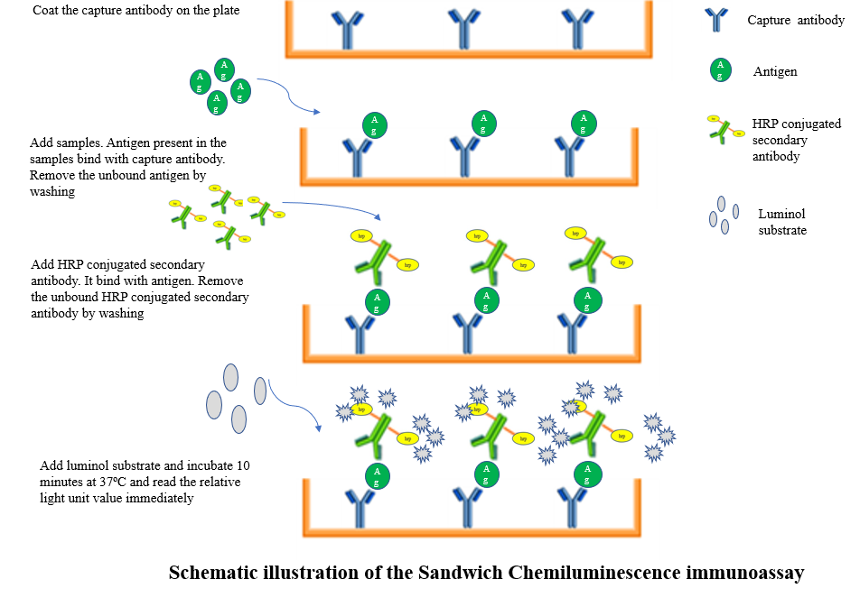
4.1 Sandwich Chemiluminescence immunoassay
1. Add 100 ul of 1ug/ml capture antibody or cell lysate or serum in the microwell strips. Seal with tape and press firmly onto top of microwells. Incubate the plate for 2 hr at room temperature. Alternatively, the plate can be incubated overnight at 4°C.
2. Discard plate contents into a receptacle.
(1)Wash 4 times with 1X Wash Buffer, 150 µl each time per well.
(2)For each wash, strike plates on fresh paper towels hard enough to remove the residual solution in each well, but do not allow wells to dry completely at any time.
(3)Clean the underside of all wells with a lint-free tissue.
3. Add 50 µl of antigen or analyte need to detect to each well. Seal with tape and incubate the plate at room temperature for 1 hr.
4. Repeat wash procedure (Step 2).
5. Add 50 µl of HRP-linked secondary antibody to each well. Seal with tape and incubate the plate at room temperature for 30 min.
6. Repeat wash procedure (Step 2).
7. Prepare detection reagent working solution by mixing equal parts 2X luminol Reagent and 2X Peroxide.
8. dd 50 µl of the detection reagent working solution to each well.
(1)Use a plate-based luminometer set at 425 nm to measure Relative Light Units (RLU) within 1–10 min following addition of the substrate. (Optimal signal intensity is achieved when read within 10 min.)
4.2 Electrochemiluminescence immunoassay
The assays will be run in MSD’s multiplexed U-PLEX format. The U-PEX format employs multi-well plate consumables, in which each well comprises a screen-printed carbon ink electrode supporting a generic 10-plex array of binding reagents selected for their specificity to a set of 10 U-PLEX "linkers".
Capture antibodies are coupled to these linkers (using reagents available from MSD) to generate reagents that are targeted to specific elements of the generic arrays, enabling the solution phase self-assembly of capture antibody arrays. Running assays in the U-PLEX format involves the following steps:
(1)incubate a mixture of up to 10 capture antibody-linker conjugates in the U-PLEX wells to self-assemble a capture antibody array and wash to remove excess unbound capture antibody.
(2)Add the sample and a sample diluent comprising blocking components (including blockers of human anti-mouse antibodies), incubating to bind analyte, and then washing to remove unbound sample.
(3)Add labeled detection antibodies and incubate to bind captured analyte and form sandwich complexes, and then washing to remove unbound detection antibody.
(4)Add an ECL read buffer (MSD Read Buffer Gold) and analyze the plate on a MSD plate reader. The plate reader applies an electrical potential to the electrode in each well and measures the resulting ECL emission from each array element on the electrode.
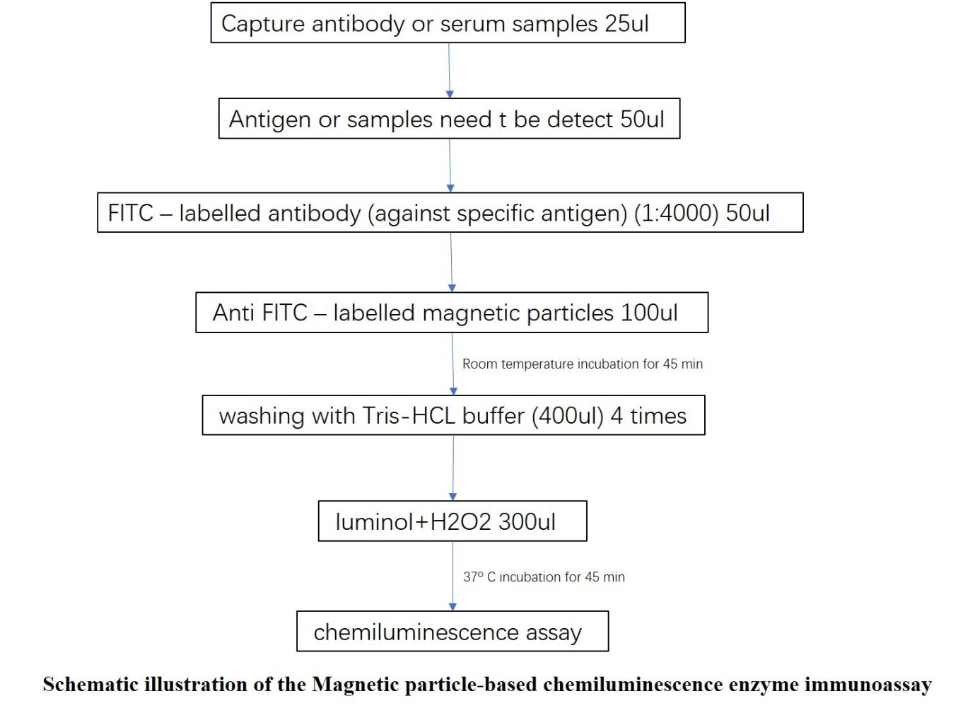
4.3 Magnetic particle-based chemiluminescence enzyme immunoassay
(1)First, add the capture antibody or serum into the test tubes.
(2)Add 50 ul of antigen or analyte (dilution ratio of 1:1000).
(3)After that, add 50 ul of FITC labeled antibody (dilution ratio of 1:4000) and 100 ul of anti-FITC antibody coated MPs. Afterwards, the mixture was incubated at room temperature for 45 min (immunoreaction time).
(4)Insert the samarium–cobalt magnet under the test tube rack for separation (5 min).
(5)The antibody coated MPs and any specific captured substances were attracted by the magnets to the bottom of the test tubes, and remove the free substances by gently tapping the test tubes against tissue paper.
(6)During the washing steps, add 400 ul of washing solution into the test tubes by placing them outside the magnet, so the MPs were resuspended in the washing solution.
(7)Finally, add 300 ul of CL substrate solution and incubate the mixture for 10 min at 37 ◦C and the emitted photons were measured.
 Guidence of GeneMedi's protocol / procedure for the diagnostics application: