


The strategies used in diagnosis of animal infectious disease for animal health
Rapid diagnosis is critical for the implementation of efficient control strategies against animal infectious and zoonotic diseases. Developing high-quality diagnostic methods and understanding animal diseases infection dynamics are important to obtain reliable diagnostic results. Here we discuss some of the most common animal disease and zoonotic disease identification methods, currently being used both in experimental and diagnostic assays.
Lateral flow assays
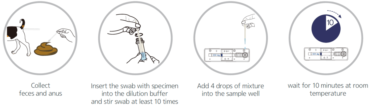
Lateral flow test, a simple cellulose-based device developed to detect the presence of a target analyte in a liquid sample [4]. There are two main variations of lateral flow tests: antigen-based test, using monoclonal antibodies to detect the specific viral antigens, another is antibody-based test, using viral antigen protein to measure the specific antibody level in a sample. For antigen-based test, test strips are coated with antibodies that bind to a viral protein and if the animal’s sample contains such proteins, they will bind to the antibodies, to form a colored indicator on the strip. Samples can be feces, eye mucus, whole blood, serum or plasma. For antibody-based test, test strips are coated with viral antigens that bind to antibodies and if the animal blood sample contains such antibodies, they will bind to the viral antigens, to form a colored indicator on the strip. Samples can be whole blood, serum or plasma [5].
Colloidal gold nanoparticles are the most widely adopted material to induce a color change when it comes in contact with the analyte. Based on the specific immune response of antigen and antibody, colloidal gold particles were used as one of the tracer markers. Driven by solvent chromatography, the markers had an immune response on the C/T line, and the detection results could be obtained according to the color of the T line. GICA samples can be whole blood, serum or plasma, and studies have shown that the colloidal gold reagent has a high consistency in detecting whole blood, plasma or serum [6]. The test results could be provided between 10-30 mins from sample collection.
Due to the low sensitivity of this test, it would effectively work only on symptomatic individuals and these tests could be less reliable in comparison with RT-PCR tests. However, it could be quickly performed at the point-of-care, or in community settings without the need for expensive equipment. An overview of the process for lateral flow assay is presented in Figure 1.
antigen-based Lateral flow test
antibody-based Lateral flow test
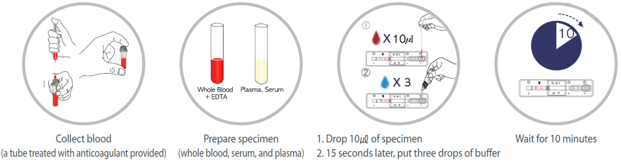
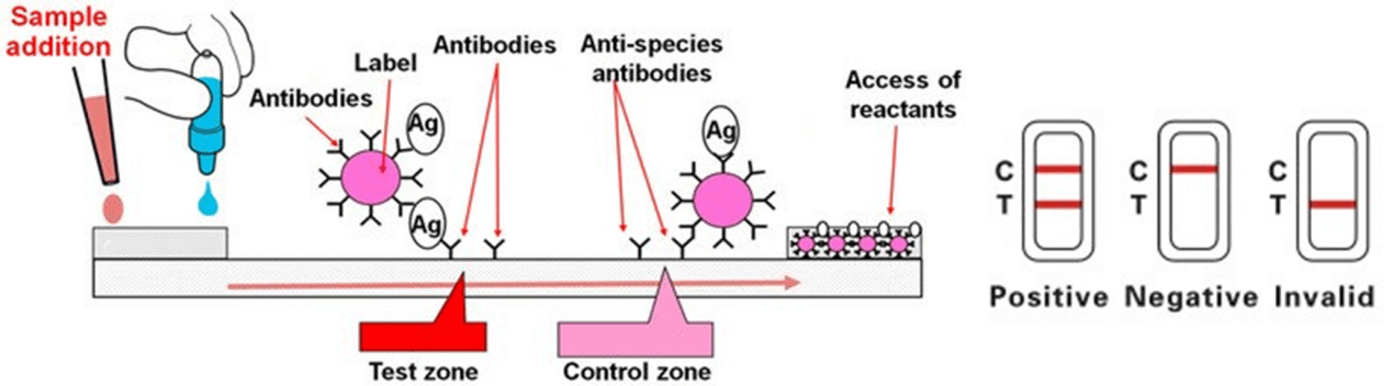
Figure 1. An overview on lateral flow assay for a serological test
Enzyme-Linked Immunosorbent Assays (ELISA)
Enzyme-linked immunosorbent assays (ELISAs) incorporate the sensitivity of simple enzyme assays with the specificity of antibodies, by employing antigens or antibodies coupled to an easily-assayed enzyme. As such ELISA is much more rapid method than immunoblotting to detect specific viral protein from a cell, tissue, organ, or body fluid. There are two main variations of ELISAs: antigen-capture ELISA (detecting viral proteins), involve attachment of a capture antibody to a solid matrix for the viral protein of interest, while antibody-capture ELISA measures the specific antibody level in a sample, by coating viral antigen protein on a solid surface.
There are two principles based on antigen-capture and antibody-capture ELISAs. In a general, ELISAs are considered a highly sensitive method that can detect a fairly low number of proteins at the range of picomolar to nanomolar range (10-12 to 10-9 moles per liter). ELISA method was found useful as a diagnostic tool to detect influenza viral antigen much quicker than other conventional virus detection methods [7]. In another previous study, comparison of ELISA, with conventional methods has demonstrated ELISA superiority for the rapid detection and identification of influenza A virus [8]. A simplified and standardized neutralization enzyme immunoassay (Nt-EIA) was developed to detect measles virus growth in Vero cells and to quantify measles neutralizing antibody [9].
Newer EIA formats for hepatitis C virus diagnostics have been constantly evaluated [10, 11]. As such ELISAs are being used for plethora of application both in experimental and diagnostic virology including dengue, and influenza [12-14]. On the other hand, although rapid than traditional plaque assays or TCID50, ELISA assays sometimes could be quite expensive, due to the cost of reagents used. Unfortunately, sometimes required antibodies may not be commercially developed as well. In contrast, attempts to develop antibodies in-house may be quite expensive. Additional variability may also be introduced due to high background signals generated by non-specific binding, or cross-reactivity with non-viral protein targets.
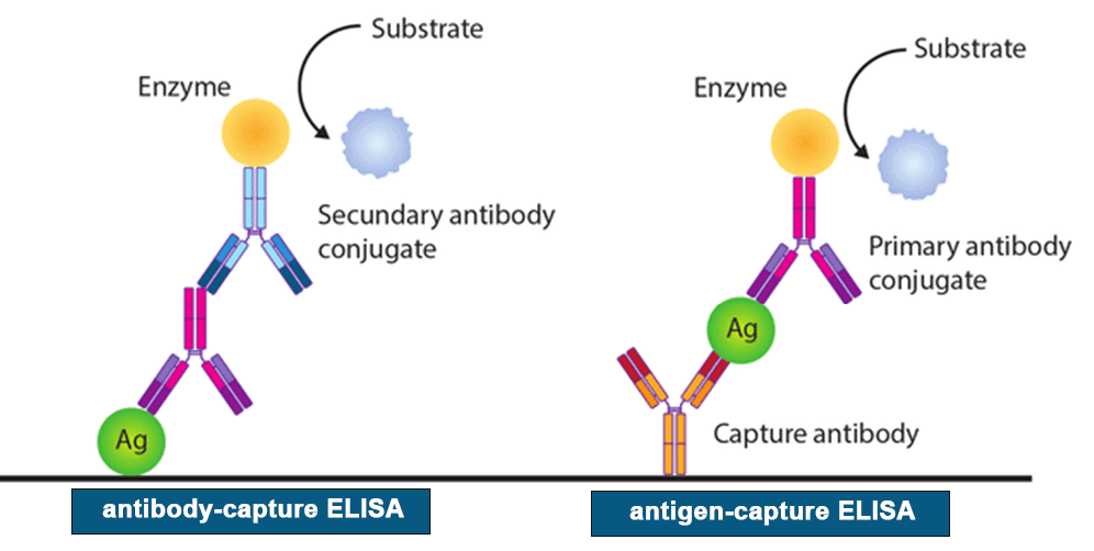
Figure 2. A schematic representation of two principles based on antigen or antibody capture ELISA [15].
Immunofluorescence staining (IF)
The immunofluorescence staining is to detect viral antigen using virus-specific monoclonal or polyclonal antibodies fluorescent staining of viral antigens is visualized under a fluorescent microscope.
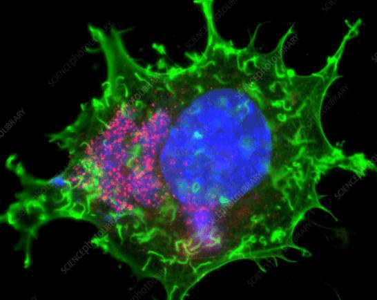
Figure 3. Immunofluorescence staining of vaccinia virus infected cell [16]. Areas of virus assembly within the cell are pink. Host and viral DNA (deoxyribonucleic acid) is blue. The host cell's DNA is contained within its nucleus (large oval). Actin protein filaments, which make up part of the cytoskeleton, are green.
Immunohistochemistry (IHC)
Immunohistochemistry is to detect viral antigen in formalin-fixed paraffin-embedded tissues using virus-specific monoclonal or polyclonal antibodies followed by an enzyme-linked secondary antibody and chemical substrate; IHC can be visualized under alight microscope.
In situ hybridization (ISH)
In situ hybridization (ISH) is to detect viral nucleic acid present in fixed tissues using a labeled complementary DNA, RNA or modified nucleic acid strand. Different with PCR approach where viral nucleic acid in a sample is amplified before detection, ISH detects viral nucleic acid that is not going through an amplification process.
Virus isolation (VI)
Obtaining the virus isolate that can efficiently grow in cell culture is critical for pathogenesis study, development of diagnostic assays, and vaccine development. However, viral culture results do not yield timely results to inform clinical management. Shell-vial tissue culture results may take 1-3 days, while traditional tissue-cell viral culture results may take 3-10 days. Due to the long incubation time, high technical requirements, and must be carried out in a level III safe biological laboratory, it is not suitable for rapid virus diagnosis during the epidemic period [17].
Electron microscopy (EM)
Electron microscopy allows direct visualization of virus particles. Two EM techniques are commonly used in diagnostic laboratories: negative-stain EM and ultrathin-section EM. Negative-stain EM for detection of virus particles in a fluid matrix; ultrathin-section EM for detection of virus particles in fixed tissues or cells. Based on characteristic morphology and size of virus particles observed under EM, viruses can be assigned to appropriate family, e.g., coronavirus-like particles were observed in some feces during initial investigation of diarrheic cases caused by porcine delta-coronavirus. Although EM cannot identify viruses to the species level, identification to the family level can still facilitate next-step testing to achieve definite diagnosis. However, EM generally is less sensitive and needs presence of sufficient amount of virus (about105–6virions per milliliter) in examined specimens. In addition, EM requires expensive equipment and highly skilled microscopist.
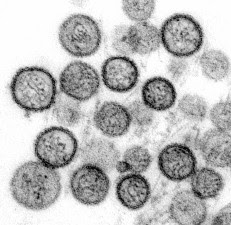
Figure 4. Transmission Electron Microscopy of hantavirus virions[18].
Molecular Methods
The development of molecular methods for the direct identification of a specific viral genome from the clinical sample is one of the greatest achievements of the 21st century. Polymerase chain reaction (PCR) is a technique that can in vitro amplify specific nucleic acid sequences and produce billion copies of target sequences within a few hours. Reverse Transcription-Polymerase Chain Reaction (RT-PCR) is proven technology leaders for rapid detection and molecular identification for most known human viruses [19]. Virus can be detected through real-time RT-PCR with primers against two segments of virus RNA genome. However, high mutation rates may lead to extensive changes in viral nucleic acid sequences making dedicated PCR primer use irrelevant, therefore there is high demand for the development of rapid and universal virus identification and detection technologies.
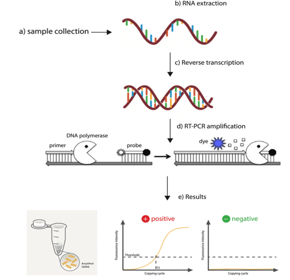
Figure 5. An overview of RT-PCR of virus detection [20].
Category
Genemedi provides diagnostic antibodies and antigens for the in vitro diagnosis of
diseases from the Companion Animal, Cat/Feline, Dog/Canine, Rabbit, Bovines/Cattle,
Ovines/Sheep, Caprine/Goat, Equine/Horse, Swine/Porcine/Pig, Avian/bird/poultry, Fish and so
on.
Animal category


Disease category
Veterinary NCDs (noncommunicable diseases)
-
· Acute inflammation, infections
· Inflammatory diseases, infections
· Myocardial injury, heart disease· Bacterial infections, sepsis
· Heart failure, cardiac disease
· Thrombosis, disseminated intravascular coagulation
Veterinary Infectious Disease
-
· Infectious bursal/Gumboro disease
· Foot and mouth disease
· Hemolytic uremic syndrome/E. coli infection· Bovine spongiform encephalopathy (BSE)
· Brucella abortus, Brucella melitensis, Brucellosis
· Monkeypox, cowpox
Technical resource
Reference:
1. Ryu, S., et al., One Health Perspectives on Emerging Public Health Threats. J Prev Med Public Health, 2017. 50(6): p. 411-414.
2. World Health Organization (WHO). Zoonoses. Accessed October 3, 2018.; Available from: http://www.who.int/zoonoses.
3. Leslie MJ, M.J. Surveillance for zoonotic diseases. BLUKO97- Mikanatha 2007; Available from: http://courses.washington.edu/zepi526/Papers08/Rabies%20chapter.pdf.
4. Available from: https://www.doctorsgate.com/en/what-are-the-current-diagnostic-tests-for-covid-19/.
5. Anylab. Available from: www.zetbio.com.
6. Li, H., et al., A new and rapid approach for detecting COVID-19 based on S1 protein fragments. Clin Transl Med, 2020. 10(2): p. e90.
7. Khanna, M., et al., Evaluation of influenza virus detection by direct enzyme immunoassay (EIA) and conventional methods in asthmatic patients. J Commun Dis, 2001. 33(3): p. 163-9.
8. Waner, J.L., et al., Comparison of Directigen FLU-A with viral isolation and direct immunofluorescence for the rapid detection and identification of influenza A virus. J Clin Microbiol, 1991. 29(3): p. 479-82.
9. Cameron, J.D., A.P. Skubitz, and L.T. Furcht, Type IV collagen and corneal epithelial adhesion and migration. Effects of type IV collagen fragments and synthetic peptides on rabbit corneal epithelial cell adhesion and migration in vitro. Invest Ophthalmol Vis Sci, 1991. 32(10): p. 2766-73.
10. Kim, M.H., S.Y. Kang, and W.I. Lee, Evaluation of a new rapid test kit to detect hepatitis C virus infection. J Virol Methods, 2013. 193(2): p. 379-82.
11. Niu, X., et al., [Establishment of the evaluation reference system for domestic anti-hepatitis C virus diagnostic enzyme immunoassay kits]. Xi Bao Yu Fen Zi Mian Yi Xue Za Zhi, 2013. 29(7): p. 761-4.
12. Filice, G., et al., Sensitivity and specificity of anti-HIV ELISA employing recombinant (p24, p66, gp120) and synthetic (gp41) viral antigenic peptides. Microbiologica, 1991. 14(3): p. 185-94.
13. de Boer, G.F., W. Back, and A.D. Osterhaus, An ELISA for detection of antibodies against influenza A nucleoprotein in humans and various animal species. Arch Virol, 1990. 115(1-2): p. 47-61.
14. Cuzzubbo, A.J., et al., Comparison of PanBio dengue duo enzyme-linked immunosorbent assay (ELISA) and MRL dengue fever virus immunoglobulin M capture ELISA for diagnosis of dengue virus infections in Southeast Asia. Clin Diagn Lab Immunol, 1999. 6(5): p. 705-12.
15. Maria-C-Jimenez-Martinez. Available from:
https://www.researchgate.net/profile/Maria-C-Jimenez-Martinez/publication/320265684/figure/fig3/AS:547016444395520@1507430291554/ELISA-assays-Direct-ELISA-mostly-used-for-antigen-detection-Indirect-ELISA-mainly-used.png.
16. sciencephoto. Available from: https://www.sciencephoto.com/media/90184/view.
17. Uyeki, T.M., et al., Clinical Practice Guidelines by the Infectious Diseases Society of America: 2018 Update on Diagnosis, Treatment, Chemoprophylaxis, and Institutional Outbreak Management of Seasonal Influenzaa. Clin Infect Dis, 2019. 68(6): p. e1-e47.
18. Goldsmith, C. Hantavirus Life Cycle and Infection Process. Available from: https://www.hantasite.com/2017/03/hantavirus-life-cycle-and-infection.html.
19. METHODS, M. 2013; 3:207
20. biotech, G.; Available from: https://www.globalbiotechinsights.com/articles/20247/the-worldwide-test-for-covid-19.
