


The pathogenesis of asthma is far more complex than the phrase "airway inflammation" suggests. Type Ⅱ inflammation, eosinophils, cytokine networks… each link influences the direction of treatment strategies. In recent years, an ancient but often overlooked player—macrophages—is re-entering researchers' field of view.
Macrophages: The "Two-Faced Sentinels" in the Lung
The lungs harbor a large number of macrophages, which serve as a bridge between innate immunity and adaptive immunity. Traditionally, macrophages are divided into alveolar macrophages (AM) and interstitial macrophages (IM), which differ in location and function.
A key question is: in the airways of asthma patients, which group of macrophages is "adding fuel to the fire"? The answer may lie in the differential expression of the surface molecule CD43. CD43-negative (CD43⁻) and CD43-positive (CD43⁺) macrophages may play distinctly different roles.
Research Background: The "Cold Case" of IL-9
Interleukin-9 (IL-9) is a pleiotropic cytokine closely associated with allergic diseases. Its levels are elevated in asthma patients, but which type of cells it acts on and through what mechanism it causes disease were previously unclear.
Researchers previously found in mouse models that IL-9-responsive macrophages are key drivers of allergic airway inflammation, and this effect depends on arginase 1 (Arg1). However, whether human macrophages follow the same pattern has always lacked direct evidence.
Research Objective: Finding the "IL-9 Target Cells" in Human Lungs
This study attempts to answer three questions: Does a specific IL-9-responsive macrophage subpopulation exist in human lungs? What changes occur in these cells in asthma patients? What downstream mechanisms are initiated upon IL-9 activation?
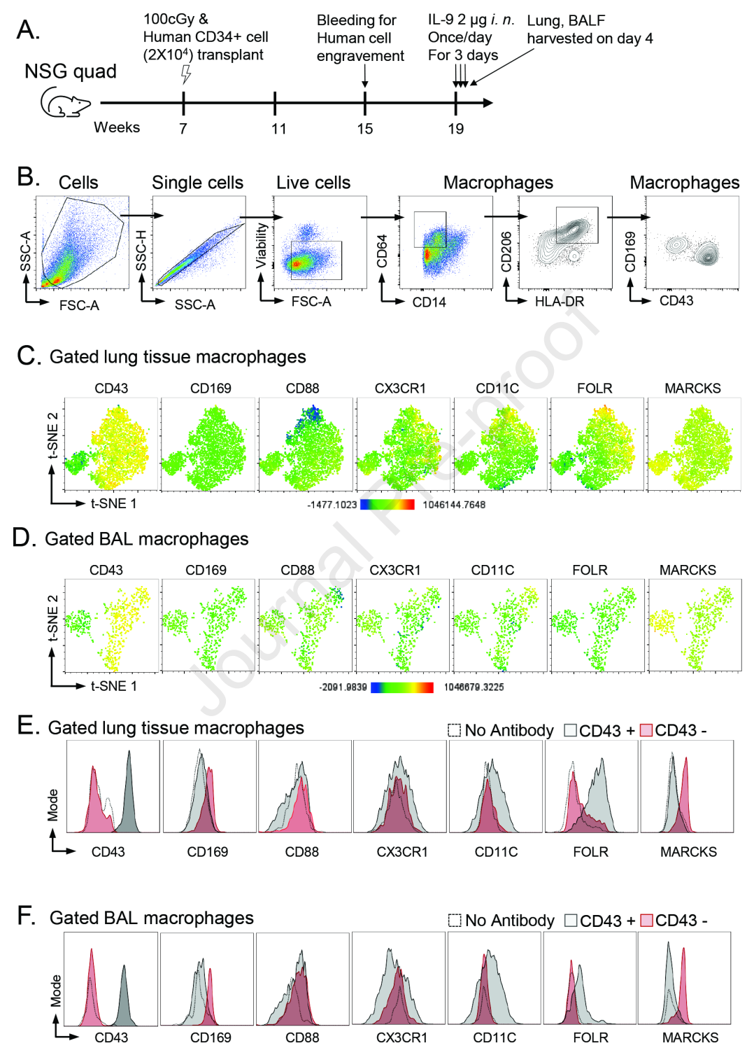
The research team adopted a dual-track strategy: on one hand, they used a humanized mouse model (NSG-Quad) to directly observe human macrophage responses to IL-9 in vivo; on the other hand, they collected bronchoalveolar lavage (BAL) samples from adult and pediatric asthma patients to verify whether the findings in animal models truly exist in humans.
Core Findings: A Complete Metabolic Pathway
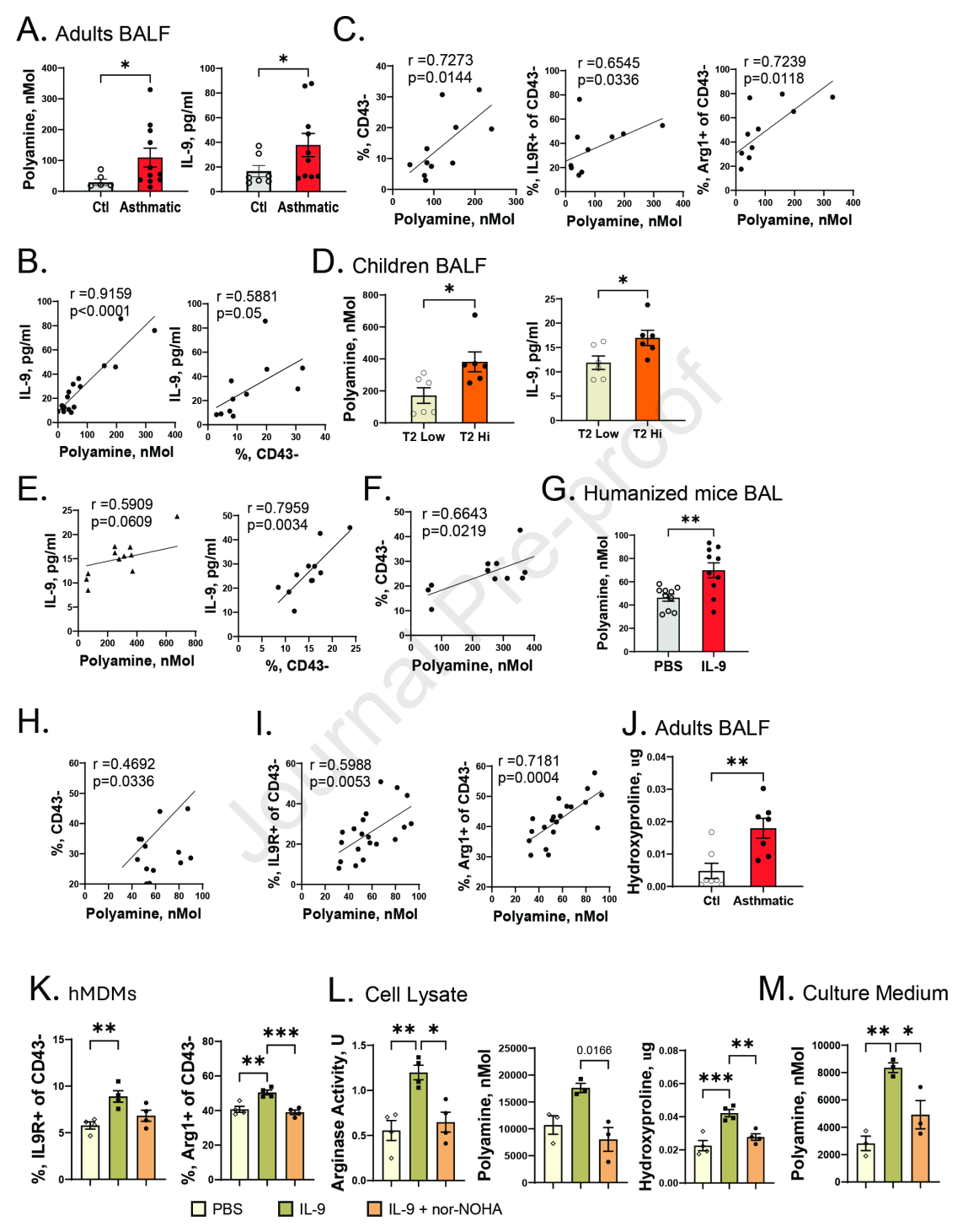
The results outline a clear chain. First, CD43⁻ macrophages are the primary target cells of IL-9. In humanized mice, three days after intranasal administration of IL-9, CD43⁻ macrophages significantly expanded; the same trend was also observed in the BAL of asthma patients.
Second, IL-9 upregulates the receptor (IL-9R) and Arg1 expression on CD43⁻ macrophages themselves, forming a positive feedback amplification effect. More critically, after Arg1 activation, it converts L-arginine into polyamines—this metabolite is significantly elevated in the BAL of asthma patients and positively correlates with IL-9 levels and the proportion of CD43⁻ macrophages.
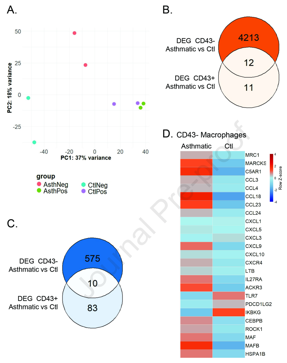
Transcriptome sequencing further revealed that CD43⁻ macrophages from asthma patients exhibit a unique gene expression profile: 4,225 genes upregulated, including chemokines such as CCL18 and CCL23, while changes in CD43⁺ macrophages were minimal. This indicates that the CD43⁻ subpopulation is the "most active molecular factory" in asthma inflammation.
Innovation and Breakthrough: Closing the Loop from Phenomenon to Mechanism
The value of this study lies in completing the closed loop from "phenomenon observation" to "mechanism verification." Researchers treated human monocyte-derived macrophages with the selective arginase inhibitor nor-NOHA and found that IL-9-induced polyamine synthesis was completely blocked, thereby confirming that polyamine production indeed depends on the Arg1 pathway.
Another highlight is the inclusion of pediatric samples. The study found that in the BAL of children with Type Ⅱ high inflammation (T2-high) asthma, the proportion of CD43⁻ macrophages and their IL-9R and Arg1 expression were higher than in T2-low children, indicating that this pathway is equally active in childhood asthma.
Practical Significance: Potential New Targets for Precision Therapy
This study provides a new dimension for explaining the heterogeneity of asthma. Not all asthma patients depend on this pathway—it is more likely to drive a subtype characterized by high IL-9 expression.
From a translational perspective, the IL-9R/Arg1/polyamine axis provides multiple potential intervention nodes for drug development: blocking the binding of IL-9 to its receptor, inhibiting Arg1 enzyme activity, or modulating the proportion of macrophage subpopulations may all become strategies for future precision therapy.
The study also suggests that polyamine concentration in BAL may serve as a biomarker for determining asthma subtypes, helping clinicians screen patients suitable for anti-IL-9 therapy.
Summary and Reflections
This study provides solid evidence for the hypothesis that the "IL-9—macrophage—metabolic reprogramming" axis drives asthma inflammation. It reminds us that the function of immune cells is not only reflected in the secretion of cytokines but also in reshaping the local metabolic environment.
A line of thought worth pondering is: polyamines play a dual role in tissue repair and remodeling—moderate amounts promote healing, while excessive amounts lead to fibrosis. Is the elevated polyamine level in the airways of asthma patients compensatory repair or the beginning of pathological remodeling? If this pathway is blocked, can inflammation be suppressed without affecting normal repair? These questions await deeper mechanistic research and clinical validation to answer.