


Immuno-Diagnostics Antibodies and Antigens for Prosthetic Joint Infection (PJI)
What is Prosthetic Joint Infection (PJI)?-Introduction
Prosthetic Joint Infection (PJI) is an infection that occurs around artificial joint implants used in joint replacement surgeries. As one of the most severe complications in orthopedic surgery, it significantly impacts patient quality of life and is both challenging and costly to treat [1-3].
Pathophysiology
Prosthetic joint infections are primarily caused by bacteria, such as Staphylococcus aureus (including methicillin-resistant MRSA strains) and coagulase-negative staphylococci, that invade the surgical site during or shortly after surgery. These pathogens form biofilms on the surface of the prosthetic joint that are highly resistant to both the immune system and antibiotics, making the treatment of the infection particularly challenging [4-7].
The main characteristics of PJI disease are:
-
Joint pain: The most common symptom of PJI is pain, which typically arises several weeks to months postoperatively, and primarily manifests as either persistent or intermittent pain.
-
Joint swelling: The affected joint typically presents with significant swelling in cases of infection.
-
Redness and warmth: The skin surrounding the joint may become red and warm during infection, which are classic signs of infection.
-
Joint effusion: In cases of infection, fluid often accumulates within the joint, and the synovial fluid is typically cloudy or purulent.
Why is the use of biomarkers important in diagnosing PJI?
-
Improved Diagnostic Accuracy: Bacterial culture and imaging are not sensitive and specific methodologies for diagnosing PJI. More accurate biomarkers (such as C-reactive protein, interleukin-6, alpha defensin) may be possible if bacterial cultures are negative but there is infection.
-
Early Detection of Infection: Biomarkers can be detected in the early stages of infection, allowing physicians to diagnose PJI before symptoms fully manifest.
-
Guiding Treatment Decisions: Biomarkers help physicians make informed treatment decisions.
-
Monitoring Treatment Effectiveness: Regular biomarker testing allows clinicians to monitor treatment progress in real time.
-
Distinguishing Between Infection Types: Biomarkers assist in distinguishing between bacterial infections and non-infectious inflammatory responses.
-
Less Invasive Diagnostics: Biomarker testing typically requires only blood or joint fluid samples.
-
Addressing a Wide Range of Pathogens: Biomarker testing is pathogen-independent.
-
Identifying Biofilm Infections: Biomarkers may be useful to identify biofilm-associated infections.
Biomarkers for the detection of PJI
Key Biomarkers in Synovial Fluid for PJI (Periprosthetic Joint Infection) and Their Pathological Concentrations
| PJI Biomarker Category | Name | Pathological Concentration in Synovial Fluid |
|---|---|---|
| Alpha-defensin | Alpha-defensin-1 | > 50 μg/L (elevated in PJI, sensitive for biofilm-associated infections) |
| Alpha-defensin | Alpha-defensin-3 | > 50 μg/L (good reference for PJI, often tested with other defensins) |
| Alpha-defensin | Alpha-defensin-5 | > 20 μg/L (antimicrobial peptide, detected in synovial fluid) |
| Inflammatory Marker | Interleukin-6 (IL-6) | / (rises rapidly after infection, useful for early detection) |
| Inflammatory Marker | C-reactive protein (CRP) | / (elevated in acute PJI but non-specific; used with other tests) |
| Inflammatory Marker | sCD14-ST/Presepsin | / (associated with monocyte activation, useful for infection diagnosis) |
Notes:
Alpha-defensins: These are key biomarkers for PJI, with defensin-1 and-3 showing high sensitivity (especially for biofilm-related infections) and defensin-5 having a role in gut defense but also being detected in synovial fluid for PJI.
IL-6: Useful for early detection of PJI (especially after surgery) as its levels rise quickly after infection onset.
CRP: Elevated in acute PJI but not specific to PJI alone; it needs to be combined with other diagnostic results.
sCD14-ST/Presepsin: Linked to monocyte activation and can help diagnose bacterial infections (including PJI).
Technologies and Platforms
| Technology | Overview | Advantages |
|---|---|---|
| ELISA (Enzyme-Linked Immunosorbent Assay) | A commonly used detection technology that quantifies specific proteins or other molecules in samples through the combination of antibodies and enzyme labels. | High sensitivity, strong specificity, simple operation, and relatively low cost. |
| Simoa (Single Molecule Array) | An ultra-sensitive detection technology that can detect biomarkers at the single-molecule level through digital readout and signal analysis. | Extremely high sensitivity and accuracy, suitable for early diagnosis and monitoring. |
| Multiplex Assays | A technology that allows the simultaneous detection of multiple biomarkers, improving detection efficiency and data throughput by using different fluorescent or chemical labels to distinguish each biomarker. | High throughput, sample and reagent savings, strong data integration. |
| ECL (Electrochemiluminescence) | A high-sensitivity detection method that combines electrochemical and luminescence technologies to analyze biomarkers and molecular interactions. | Extremely high sensitivity and specificity, low background noise, suitable for detecting low-concentration samples and high-throughput screening. |
| LC-MS/MS (Liquid Chromatography-Tandem Mass Spectrometry) | A high-precision detection method that combines liquid chromatography and mass spectrometry to analyze compounds in complex biological samples. | High sensitivity and specificity, capable of detecting multiple target molecules simultaneously, suitable for metabolomics and proteomics research. |
Clinical Applications and Research
| Name | Sample | Biomarkers | Method |
|---|---|---|---|
| Zimmer Biomet | Synovasure | Alpha-defensin 1 | LFIA |
| HansaBioMed Life Sciences | Blood/Synovasure | Exosome levels | ELISA |
Specificity Validation data on defensin-1/defensin-5 protein
GeneMedi's alpha defensin-1 antibodies exhibit excellent specificity, and sensitivity in ELISA with the best results achieved by using GMP-h-a defensin-1-Ab05 for coating and Ab04 for detection. GeneMedi's GMP-h-a-defensin-1-Ab04 and GMP-h-a-defensin-1-Ab05 antibodies demonstrate superior specificity in direct ELISA assays, as they can bind a-defensin-1 and do not bind a-defensin-5. In the sandwich ELISA experiment, coating with GMP-h-a-defensin-1-Ab05 and detecting with GMP-h-a-defensin-1-Ab04 yields better results than coating with GMP-h-a-defensin-1-Ab04 and detecting with GMP-h-a-defensin-1-Ab05, indicating that the choice of antibodies and their order of use have a significant impact on the experimental outcomes.
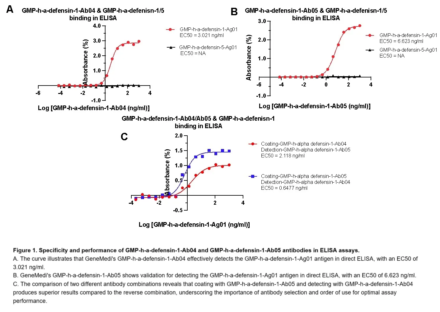
Figure 1. Specificity and performance of GMP-h-a-defensin-1-Ab04 and GMP-h-a-defensin-1-Ab05 antibodies in ELISA assays.
- A. The curve illustrates that GeneMedi's GMP-h-a-defensin-1-Ab04 effectively detects the GMP-h-a-defensin-1-Ag01 antigen in direct ELISA, with an EC50 of 3.021 ng/ml.
- B. GeneMedi's GMP-h-a-defensin-1-Ab05 shows validation for detecting the GMP-h-a-defensin-1-Ag01 antigen in direct ELISA, with an EC50 of 6.623 ng/ml.
- C. The comparison of two different antibody combinations reveals that coating with GMP-h-a-defensin-1-Ab05 and detecting with GMP-h-a-defensin-1-Ab04 produces superior results compared to the reverse combination, underscoring the importance of antibody selection and order of use for optimal assay performance.
The stability of GeneMedi's Alpha defensin-5 antigen is significantly improved after adding GeneMedi's buffer 2. The stability of GeneMedi's alpha defensin-5 antigen when stored in phosphate-buffered saline (PBS) is relatively poor, which can pose challenges for reliable storage and usage in various applications. Initial assessments reveal that this antigen exhibits instability at room temperature as well as at low temperatures, regardless of whether it is in lyophilized or liquid form. This can lead to degradation or loss of biological activity, raising concerns for researchers relying on consistent performance in experiments. However, significant improvements in stability can be achieved by substituting PBS with GeneMedi's Buffer 2. This proprietary buffer not only enhances the preservation of alpha defensin-5 but also effectively mitigates the adverse effects of temperature fluctuations and physical state. With GeneMedi's Buffer 2, the alpha defensin-5 antigen maintains its structural integrity and biological function, ensuring reliable results in subsequent assays.
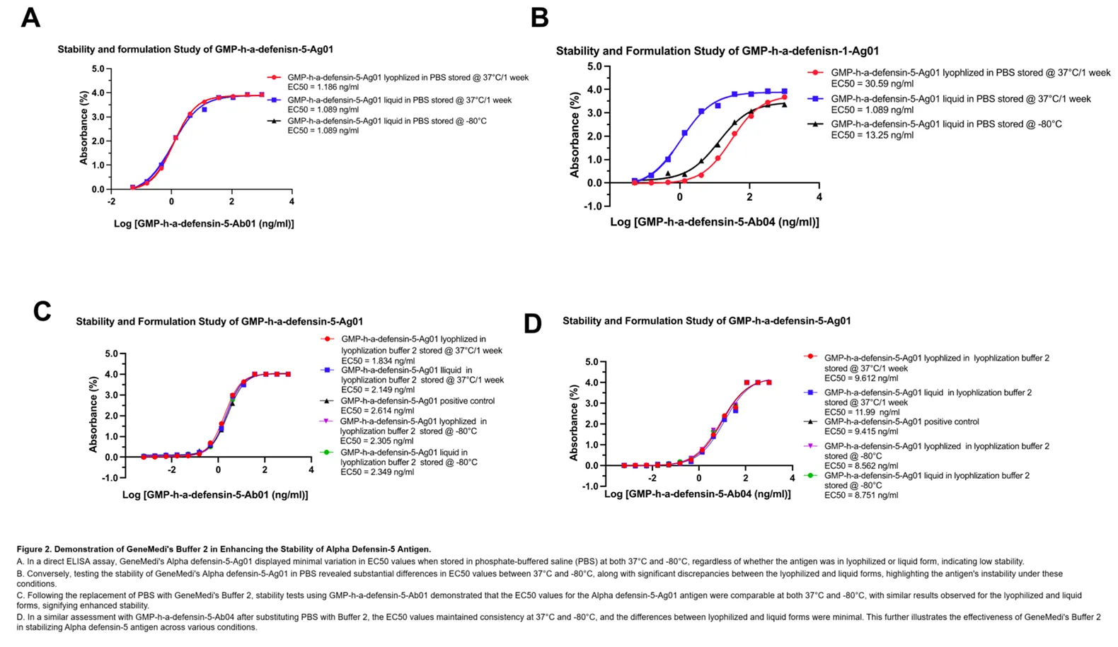
Figure 2. Demonstration of GeneMedi's Buffer 2 in Enhancing the Stability of Alpha Defensin-5 Antigen.
- A. In a direct ELISA assay, GeneMedi's Alpha defensin-5-Ag01 displayed minimal variation in EC50 values when stored in phosphate-buffered saline (PBS) at both 37°C and -80°C, regardless of whether the antigen was in lyophilized or liquid form, indicating low stability.
- B. Conversely, testing the stability of GeneMedi's Alpha defensin-5-Ag01 in PBS revealed substantial differences in EC50 values between 37°C and -80°C, along with significant discrepancies between the lyophilized and liquid forms, highlighting the antigen's instability under these conditions.
- C. Following the replacement of PBS with GeneMedi's Buffer 2, stability tests using GMP-h-a-defensin-5-Ab01 demonstrated that the EC50 values for the Alpha defensin-5-Ag01 antigen were comparable at both 37°C and -80°C, with similar results observed for the lyophilized and liquid forms, signifying enhanced stability.
- D. In a similar assessment with GMP-h-a-defensin-5-Ab04 after substituting PBS with Buffer 2, the EC50 values maintained consistency at 37°C and -80°C, and the differences between lyophilized and liquid forms were minimal. This further illustrates the effectiveness of GeneMedi's Buffer 2 in stabilizing Alpha defensin-5 antigen across various conditions.
GeneMedi's Alpha defensin-5 antibodies' stability is notably enhanced upon the addition of GeneMedi's buffer 2. GeneMedi's alpha defensin-5 antibodies, specifically GMP-h-a-defensin-5-Ab01 and GMP-h-a-defensin-5-Ab04, initially demonstrate relatively poor stability when stored in phosphate-buffered saline (PBS). Notably, this stability issue is markedly improved with the addition of GeneMedi's proprietary Buffer 2, which has been designed to optimize the preservation of biologically active compounds. During ELISA assays conducted with antibodies in PBS, the stability of GMP-h-a-defensin-5-Ab01 and GMP-h-a-defensin-5-Ab04 is significantly affected by both temperature and the physical state of the antibodies. Under varying temperature conditions, these antibodies exhibited considerable fluctuations in stability, leading to inconsistent performance in the assays. Furthermore, there was a distinct difference in stability between the lyophilized and liquid forms, adding another layer of complexity to their usage in experimental settings. However, upon transitioning from PBS to GeneMedi's Buffer 2, the improvement in antibody stability was profound. The antibodies maintained their structural integrity and biological activity regardless of the temperature extremes—whether subjected to high or low temperatures—or the form they were in (lyophilized or liquid). This significant enhancement means that temperature variations and the physical state of the antibodies no longer exert a detrimental influence on their stability.
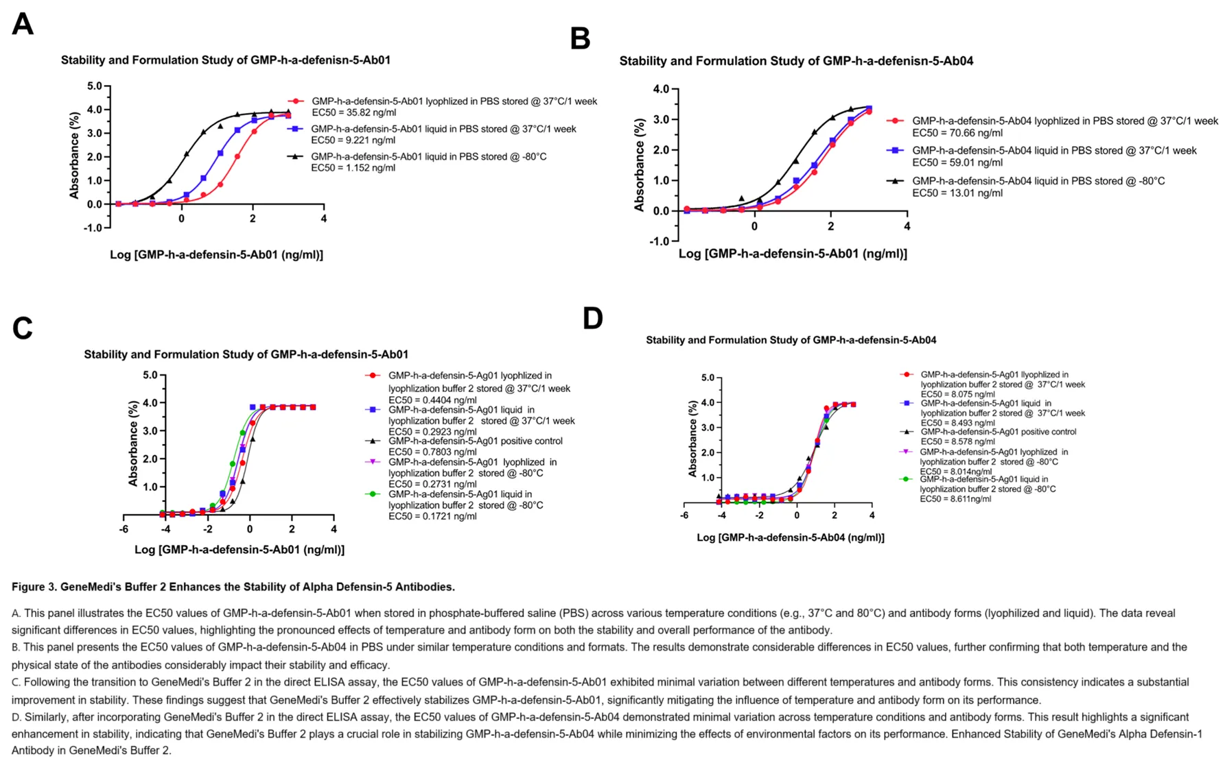
Figure 3. GeneMedi's Buffer 2 Enhances the Stability of Alpha Defensin-5 Antibodies.
- A. This panel illustrates the EC50 values of GMP-h-a-defensin-5-Ab01 when stored in phosphate-buffered saline (PBS) across various temperature conditions (e.g., 37°C and 80°C) and antibody forms (lyophilized and liquid). The data reveal significant differences in EC50 values, highlighting the pronounced effects of temperature and antibody form on both the stability and overall performance of the antibody.
- B. This panel presents the EC50 values of GMP-h-a-defensin-5-Ab04 in PBS under similar temperature conditions and formats. The results demonstrate considerable differences in EC50 values, further confirming that both temperature and the physical state of the antibodies considerably impact their stability and efficacy.
- C. Following the transition to GeneMedi's Buffer 2 in the direct ELISA assay, the EC50 values of GMP-h-a-defensin-5-Ab01 exhibited minimal variation between different temperatures and antibody forms. This consistency indicates a substantial improvement in stability. These findings suggest that GeneMedi's Buffer 2 effectively stabilizes GMP-h-a-defensin-5-Ab01, significantly mitigating the influence of temperature and antibody form on its performance.
- D. Similarly, after incorporating GeneMedi's Buffer 2 in the direct ELISA assay, the EC50 values of GMP-h-a-defensin-5-Ab04 demonstrated minimal variation across temperature conditions and antibody forms. This result highlights a significant enhancement in stability, indicating that GeneMedi's Buffer 2 plays a crucial role in stabilizing GMP-h-a-defensin-5-Ab04 while minimizing the effects of environmental factors on its performance. Enhanced Stability of GeneMedi's Alpha Defensin-1 Antibody in GeneMedi's Buffer 2.
GeneMedi's buffer 2 can improve the stability of GeneMedi's Alpha defensin-1 antibodies. GeneMedi's Alpha defensin-1 antibody has previously demonstrated poor stability when stored in phosphate-buffered saline (PBS), posing challenges for researchers seeking reliable performance in various applications. However, significant strides have been made in stabilizing this antibody by incorporating GeneMedi's proprietary Buffer 2, which has been specifically formulated to enhance the stability of biologically active compounds. The stability of GMP-h-a-defensin-1-Ab04 and GMP-h-a-defensin-1-Ab05 antibodies in PBS during ELISA assessments is heavily influenced by both temperature variations and the physical state of the antibodies. Comprehensive evaluations reveal that these antibodies exhibit considerable variability in stability under different temperature conditions. For instance, significant differences are noted in stability metrics when comparing the lyophilized form to its liquid counterpart, highlighting the challenges posed by traditional storage methods. However, the introduction of GeneMedi's Buffer 2 marks a pivotal improvement in the stability of these antibodies. After transitioning from PBS to this optimized buffer, both GMP-h-a-defensin-1-Ab04 and GMP-h-a-defensin-1-Ab05 reflected a remarkable enhancement in overall stability. Regardless of the temperature—be it high or low—or the antibody form, whether lyophilized or liquid, the antibodies maintained their structural integrity and functional efficacy.
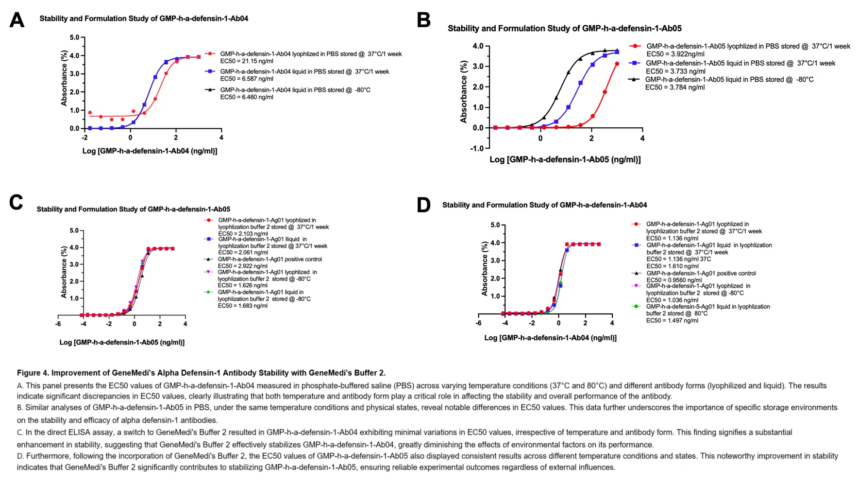
Figure 4. Improvement of GeneMedi's Alpha Defensin-1 Antibody Stability with GeneMedi's Buffer 2.
- A. This panel presents the EC50 values of GMP-h-a-defensin-1-Ab04 measured in phosphate-buffered saline (PBS) across varying temperature conditions (37°C and 80°C) and different antibody forms (lyophilized and liquid). The results indicate significant discrepancies in EC50 values, clearly illustrating that both temperature and antibody form play a critical role in affecting the stability and overall performance of the antibody.
- B. Similar analyses of GMP-h-a-defensin-1-Ab05 in PBS, under the same temperature conditions and physical states, reveal notable differences in EC50 values. This data further underscores the importance of specific storage environments on the stability and efficacy of alpha defensin-1 antibodies.
- C. In the direct ELISA assay, a switch to GeneMedi's Buffer 2 resulted in GMP-h-a-defensin-1-Ab04 exhibiting minimal variations in EC50 values, irrespective of temperature and antibody form. This finding signifies a substantial enhancement in stability, suggesting that GeneMedi's Buffer 2 effectively stabilizes GMP-h-a-defensin-1-Ab04, greatly diminishing the effects of environmental factors on its performance.
- D. Furthermore, following the incorporation of GeneMedi's Buffer 2, the EC50 values of GMP-h-a-defensin-1-Ab05 also displayed consistent results across different temperature conditions and states. This noteworthy improvement in stability indicates that GeneMedi's Buffer 2 significantly contributes to stabilizing GMP-h-a-defensin-1-Ab05, ensuring reliable experimental outcomes regardless of external influences.
Associated products
- Recommend Pair Antibody and Antigen
- Associated products
| Pair Antibody and Antigen | Products detail | Cat.No | Product Name |
| alpha defensin 1 | 1. Antigen | 1. GMP-h-a-defensin-1-Ag01 | Peptide Human alpha defensin 1 (α-defensin 1) protein |
| 2. Capture antibody | 2. GMP-h-a-defensin-1-Ab04/06 | Anti-human alpha defensin 1 (α-defensin 1) human monoclonal antibody (mAb) | |
| 3. Detector antibody | 3. GMP-h-a-defensin-1-Ab05/03 | Anti-human alpha defensin 1 (α-defensin 1) human monoclonal antibody (mAb) | |
| alpha defensin 5 | 1. Antigen | 1. GMP-h-a-defensin-5-Ag01 | Peptide Human alpha defensin 5 (α-defensin 5) protein |
| 2. Capture antibody | 2. GMP-h-a-defensin-5-Ab01/03 | Anti-human alpha defensin 5 (α-defensin 5) mouse/human monoclonal antibody (mAb) | |
| 3. Detector antibody | 3. GMP-h-a-defensin-5-Ab02/04 | Anti-human alpha defensin 5 (α-defensin 5) mouse/human monoclonal antibody (mAb) |
| Catalog No. | Products Name | Products Information |
| GMP-h-a-defensin-1 | Human alpha defensin 1 (α-defensin 1) antigen/antibody | Details |
| GMP-h-a-defensin-3 | Human Alpha-Defensin 3 (α-defensin 3) antigen/antibody | Details |
| GMP-h-a-defensin-5 | Human alpha defensin 5 (α-defensin 5) antigen/antibody | Details |
| GMP-h-IL-6 | Human Interleukin 6 (IL-6) antigen/antibody | Details |
| GMP-h-CRP | Human C-Reactive Protein (CRP) antigen/antibody | Details |
| GMP-h-a-sCD14-ST | Human sCD14-ST antigen/antibody | Details |
| GMP-h-PINP | Human PINP antigen/antibody | Details |
| GMP-h-N-MID-OC | Human N-terminal midfragment of Osteocalcin (N-MID OC) antigen/antibody | Details |
| GMP-h-PTH | Human parathyroid hormone (PTH) antigen/antibody | Details |
| GMP-SMT-25-OH-VD3/2 | Human 25 hydroxyvitamin D (25-OH-(VD3+VD2)) antigen/antibody | Details |
| GMP-SMT-25-OH-VD-3 | Human 25-hydroxy (OH) Vitamin D3 (25-OH-VD-3) antigen/antibody | Details |
| GMP-SMT-1-25-OH-VD3 | Human 1,25-hydroxy (OH) Vitamin 3 (1,25-OH-VD3) antigen/antibody | Details |
References
[1] Tande, Aaron J., and Robin Patel. "Prosthetic joint infection." Clinical microbiology reviews 27.2 (2014): 302-345.
[2] Zardi, Enrico Maria, and Francesco Franceschi. "Prosthetic joint infection. A relevant public health issue." Journal of infection and public health 13.12 (2020): 1888-1891.
[3] Pellegrini, Antonio and Virginia Suardi, "Classification and management options for prosthetic joint infection." Annals of Joint 7 (2022).
[4] Wildeman, Peter, et al. "Genomic characterization and outcome of prosthetic joint infections caused by Staphylococcus aureus." Scientific reports 10.1 (2020): 5938.
[5] Gbejuade, Herbert O. and Andrew M. "The role of microbial biofilms in prosthetic joint infections: A review." Acta orthopaedica 86.2 (2015): 147-158.
[6] Chalmers, Sarah J., and Mark E. Wylam. "Methicillin-resistant Staphylococcus aureus infection and treatment options." Methicillin-Resistant Staphylococcus Aureus (MRSA) Protocols: Cutting-Edge Technologies and Advancements (2020): 229-251.
[7] Lamret, Fabien, et al. "Antibiotic tolerance of Staphylococcus aureus biofilm in periprosthetic joint infections and antibiofilm strategies." Antibiotics 9.9 (2020): 547.
