


Tumor immunotherapy does not always work, and one often-overlooked reason lies at the starting point of the immune response. Dendritic cells (DCs) are responsible for presenting tumor information to T cells, but not all DCs are up to this task. A study published in Immunity in 2026 revealed the central role of one key class of these cells—CCR7-positive activated DCs (actDCs)—in tumor control.
Research Background: The "Division-of-Labor Confusion" of the DC Family
Traditionally, dendritic cells have been divided into two major functional subsets. cDC1 excels at cross-presentation and can efficiently activate cytotoxic CD8+ T cells; the role of cDC2 is relatively ambiguous. In recent years, single-cell sequencing has revealed that within tumors there also exists a group of DCs with a unique transcriptional signature, which highly express CCR7 and various costimulatory molecules.
This class of cells has been given multiple names: mregDC, mature DC, migratory DC. They simultaneously express activation molecules (CD80, CD86) and inhibitory molecules (PD-L1), and whether their function is pro-tumor or anti-tumor has long been debated in the field.
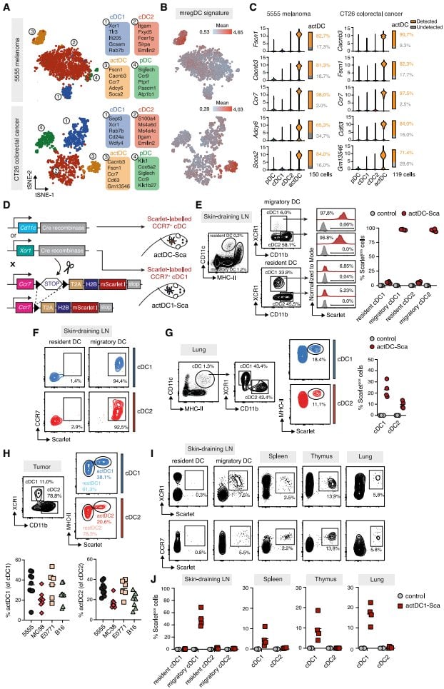
The research bottleneck lies in the fact that these cells are scarce and lack specific markers. Markers that distinguish the activated state from the resting state are also often expressed on other immune cells. Without precise tools, functional studies are impossible.
Research Objective: Fitting actDCs with a "Tracker" and a "Switch"
The core goal of this study was to develop genetic tools capable of specifically labeling and depleting actDCs. The researchers used the CCR7 gene as an entry point—it is almost universally expressed on actDCs, while absent on other DC subsets.
They constructed two types of mouse models. One inserted a fluorescent protein at the end of the CCR7 gene, enabling visual tracking of actDCs; the other inserted a diphtheria toxin receptor, allowing acute depletion of these cells at specific time points. Through different Cre driver lines, it is also possible to distinguish whether actDCs originate from cDC1 or cDC2.
Core Finding One: Only Activated-State DCs Can Prime T Cells
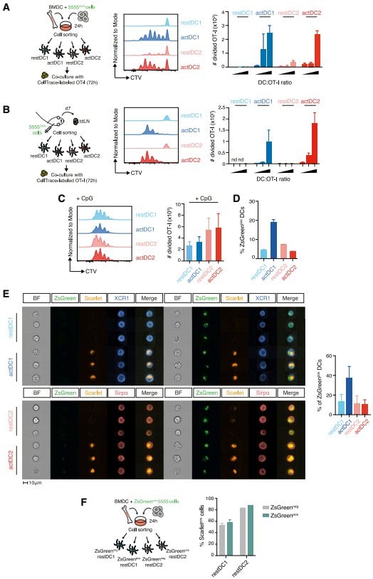
Using the above tools, the researchers divided DCs into four populations: resting cDC1, activated cDC1, resting cDC2, activated cDC2. Functional experiments gave a clear answer: only activated-state DCs can effectively activate tumor-specific CD8+ T cells.
Whether from in vitro co-culture or tumor-draining lymph nodes, resting-state DCs were unable to initiate T cell proliferation. This difference did not stem from antigen uptake capacity—all four populations could phagocytose tumor material. The key lies in whether they enter an activated state.
Core Finding Two: Two Distinct Antigen Presentation Pathways
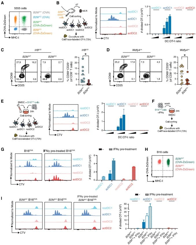
More refined mechanistic analysis revealed an unexpected division of labor. Activated cDC1 process antigens through the classical cross-presentation pathway; activated cDC2 rely on a less-noticed mechanism—cross-dressing, that is, directly acquiring pre-formed peptide-MHC complexes from the surface of tumor cells.
This difference is tumor-type dependent. In melanoma models, whether the cross-dressing function of cDC2 can be exerted depends on the abundance of MHC-I molecules on the surface of tumor cells. Interferon pretreatment can upregulate MHC-I, thereby restoring the activation capacity of cDC2.
Core Finding Three: Depleting actDCs Renders Immunotherapy Ineffective
In vivo depletion experiments confirmed the necessity of actDCs. After depleting these cells, immunogenic tumors were no longer spontaneously rejected; combined immune checkpoint inhibitors also lost efficacy, with approximately forty percent of mice showing tumor progression.
Even more noteworthy is adoptive T cell therapy. Even when cytotoxic T cells pre-activated in vitro were infused, depleting actDCs still significantly weakened efficacy. The residual T cells exhibited more severe exhaustion features—elevated expression of inhibitory molecules such as PD-1, LAG3, and TOX.
This indicates that actDCs are not only responsible for initiating the immune response, but also maintain the functional state of T cells within the tumor microenvironment, preventing their premature exhaustion.
Innovations and Significance
The breakthrough of this study lies at the tool level. The CCR7 conditional reporter and depletion mice provide a previously missing precise platform for the study of DC activation states. The study demonstrates for the first time that what determines whether T cells can be activated is not the lineage affiliation of the DC, but whether it enters an activated state.
The implications for translational research are equally clear. The state and number of actDCs may become biomarkers for predicting immunotherapy efficacy. How to induce DCs within tumors to enter an activated state may become a new strategy for enhancing existing therapies.
Summary and Reflections
actDCs stand at the hub of the tumor immune response: they are both a necessary condition for naive T cell activation and a key factor for maintaining effector T cell function. From spontaneous tumor control to immune checkpoint inhibition, and further to adoptive cell therapy, this cellular state runs throughout.
An open question worth pondering: given that actDCs simultaneously express activation and inhibitory molecules, why is their function overall biased toward immune activation? In the tumor microenvironment, which factors determine whether actDCs move toward a "pro-inflammatory" or "anti-inflammatory" balance? Understanding this regulatory logic may open a new window for next-generation combination immunotherapy strategies.