


Research Background
Severe inflammatory lung disease, including acute respiratory distress syndrome (ARDS), has a high mortality rate and limited targeted therapeutic options. Recent research suggests that inborn errors of immunity (IEI) underlie some cases. SHP1, encoded by PTPN6, is a protein tyrosine phosphatase expressed primarily in hematopoietic cells that acts as a signaling "brake" in various immune cells. SHP1 deficiency in mice leads to multi-organ immune-mediated damage. However, direct associations between PTPN6 variants and human disease had previously been reported only sporadically.
Research Objective
This study aimed to determine whether biallelic loss-of-function (LoF) variants in PTPN6 cause human disease, and to characterize the clinical phenotype and molecular mechanisms. The researchers enrolled seven affected children from five unrelated families.
Research Methods (Brief)
The study combined multiple approaches: exome sequencing of five families to identify PTPN6 variants; molecular dynamics simulations to predict the impact of variants on SHP1 structure; expression of wild-type and mutant SHP1 in COS-1 and HEK293T cells to assess protein stability, phosphatase activity, and regulation of ERK signaling; and RNA sequencing of whole blood from one patient, compared with pediatric cohorts of septic shock and ARDS.
Key Findings
All seven affected children presented with infantile-onset anemia and severe inflammatory lung disease, and most died in early childhood. Specifically:
- Patient A (II-1) developed hemolytic anemia at 4 months of age, was diagnosed with mycobacterial infection at age 13, and died of severe ARDS at age 14; autopsy showed pulmonary hemorrhage, aspergilloma, and suppurative pneumonia (Figure 1).
- Patient D (II-2) developed anemia, neutrophilic dermatitis, and interstitial lung disease at 8 months of age.
- Patient E (II-1) developed anemia and hyperbilirubinemia at 3 days of life and died of respiratory failure at 5 months of age.
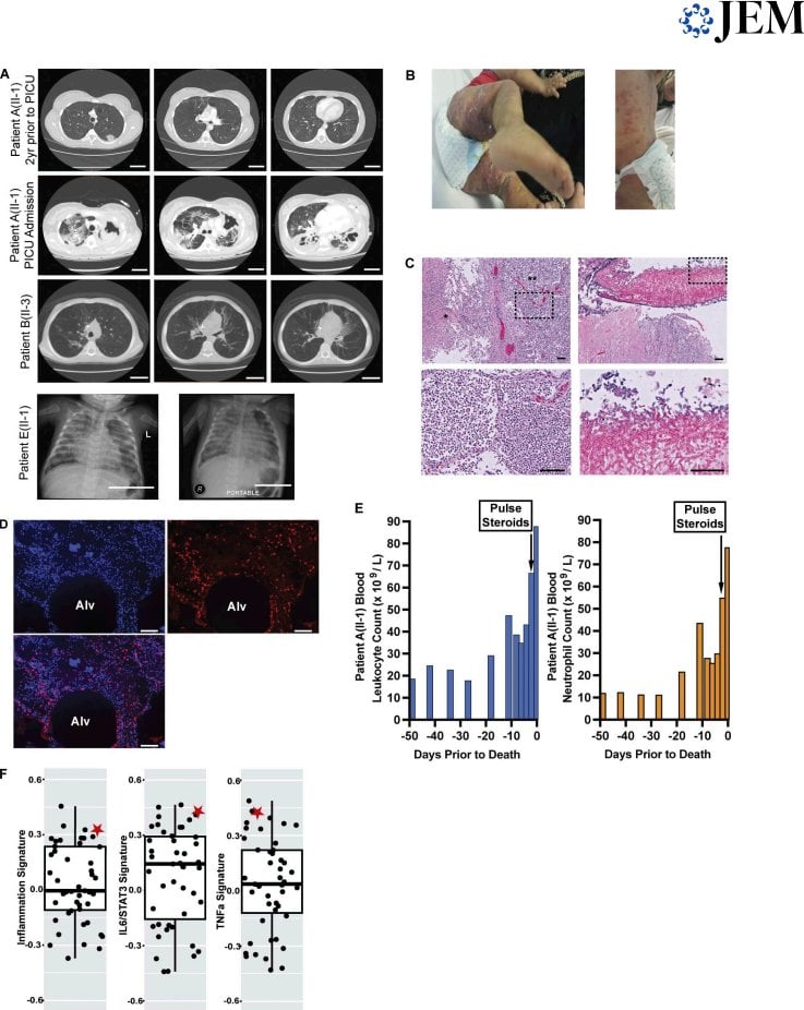
Immunological evaluation showed that some patients had low B cell counts and elevated IgA, suggesting immune dysregulation. RNA sequencing showed that inflammatory signaling pathways were significantly activated in Patient A (II-1); among a cohort of 47 septic children, this patient's inflammatory signature ranked 4th, with IL6/JAK/STAT3 and TNF/NF-κB signaling each ranking 3rd.
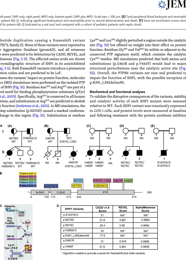
Sequencing revealed that all seven patients carried compound heterozygous or homozygous PTPN6 variants, including missense, in-frame insertion-deletion, and truncating variants. None of these variants had been reported in the gnomAD database, and all were predicted to be deleterious by multiple algorithms. The variant sites were distributed in the PTP domain or the linker region.
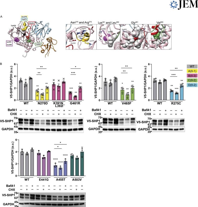
Functional experiments showed that protein levels of the SHP1N270D, SHP1G461R, SHP1V465F, and SHP1R275C mutants were significantly reduced; treatment with bafilomycin A1 restored their levels, suggesting that misfolding leads to autophagic/lysosomal degradation. Catalytic activity assays showed that SHP1G461R and SHP1V465F were completely inactive, SHP1N270D activity was markedly reduced, and SHP1R275C and SHP1K391_L392delinsNI activity was mildly reduced.
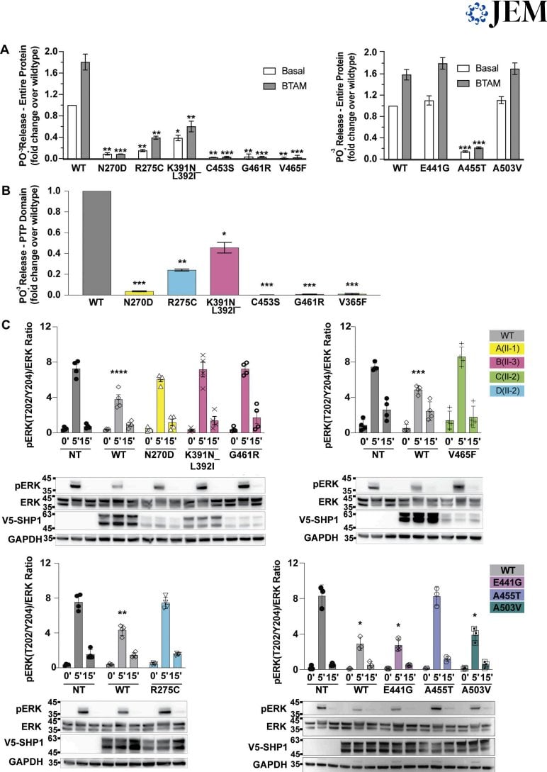
In EGF stimulation experiments, all variants associated with the ARDS phenotype failed to downregulate pERK signaling, whereas the previously reported p.E441G and the gnomAD p.A503V variants behaved similarly to wild-type. This suggests that even variants that only mildly reduce catalytic activity may still cause severe disease.
Innovations or Breakthroughs
This study established for the first time a previously unrecognized monogenic disease caused by biallelic LoF variants in PTPN6, characterized by infantile anemia and severe inflammatory lung disease. The study proposes that the severity of SHP1 dysfunction constitutes a continuous spectrum—from complete inactivation to mild functional impairment, all of which can be pathogenic. Furthermore, the study links hemolytic anemia to the CD47-SIRPα signaling axis: SHP1 participates in transmitting the "don't eat me" signal, and its deficiency can lead to antibody-independent hemolysis.
Practical Implications
This discovery expands the genetic landscape of inflammatory lung disease, suggesting that SHP1-related pathways may be potential therapeutic targets for immune-mediated lung injury. For clinicians, when encountering unexplained infantile hemolytic anemia combined with severe lung disease, the possibility of PTPN6-related disease should be considered. For translational research, SHP1 activation strategies warrant further exploration in SHP1-related lung disease and even in broader neutrophil-driven lung injury.
Summary and Reflections
This study advances SHP1 from a laboratory mouse model to a human disease entity, providing a human genetics framework for understanding the relationship between immune regulation, lung injury, and red blood cell clearance. One question worth pondering is: when the activity of a phosphatase is only mildly reduced, why is it still sufficient to trigger such a severe clinical phenotype? Does this mean that SHP1 has an irreplaceable role in specific cell types or at specific signaling nodes?