


A study published in the Journal of Experimental Medicine found that propagatable α-synuclein aggregates are present in both the cerebrospinal fluid and the dura mater of patients with Parkinson's disease; experiments in mice showed that after these aggregates deposit in brain border tissues, they activate immunity, elevate TNF, and selectively impair the drainage of macromolecules through meningeal lymphatic vessels. The study suggests that brain border tissues may actively participate in the clearance impairment of Parkinson's disease.
Research Background: Why Clearance Pathways Deserve Attention
The central nervous system lacks conventional lymphatic vessels, yet it is not without waste-removal channels. After exchange with the cerebrospinal fluid, brain interstitial fluid can drain via meningeal lymphatic vessels into the cervical lymph nodes. This pathway was long regarded as a "sewer," and only in recent years has it been re-examined.
The pathological hallmark of Parkinson's disease is the abnormal aggregation of α-synuclein (α-syn). Past research focused mostly on neuronal damage within the brain parenchyma, but aggregates also appear in peripheral compartments such as the cerebrospinal fluid, gut, and blood. α-Syn in the cerebrospinal fluid has been used to aid diagnosis, but what it does there has remained unclear.
The dura mater lies adjacent to the subarachnoid space and harbors lymphatic vessels and multiple types of immune cells, making it a crossroads of cerebrospinal fluid outflow and immune surveillance. If α-syn in the cerebrospinal fluid can act directly on this site, it would be more than a pathological marker—it could be a driver of clearance impairment.
Research Objective: Are Brain Borders a Target of Action?
The authors posed a clear question: do extracellular α-syn aggregates act directly on brain border tissues, impairing lymphatic drainage and immune homeostasis?
The study also sought to confirm three things: whether α-syn deposits are present in the dura mater of patients with Parkinson's disease; whether cerebrospinal fluid-derived α-syn can cause measurable clearance impairment in animals; and what the underlying molecular mediator is.
Research Methods (Brief): Human Tissue, Two Mouse Models, and In Vitro Cells
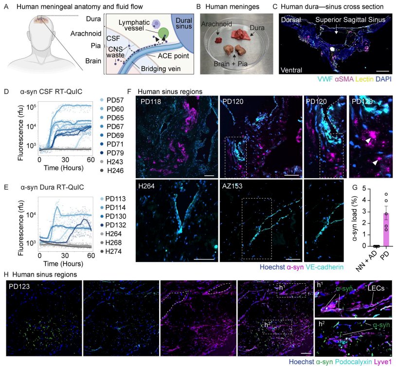
The study comprised three tiers of evidence. The first tier was postmortem human brain tissue: cerebrospinal fluid and dura mater around the superior sagittal sinus were obtained from patients with Parkinson's disease and neurologically normal controls, RT-QuIC was used to detect seeding activity, and immunohistochemistry was used to localize α-syn inclusions.
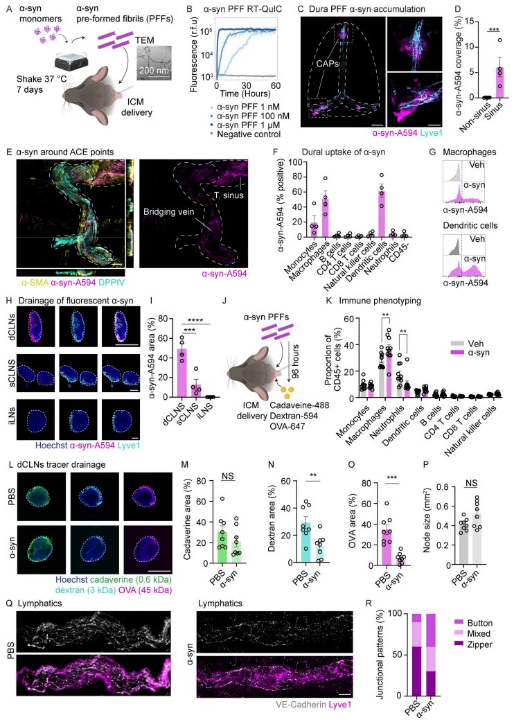
The second tier involved two mouse models. In the acute model, α-syn preformed fibrils (PFF) were injected into the mouse cisterna magna, and tissue distribution and drainage changes were observed from 24 to 96 hours; in the chronic model, an AAV9 vector was used to make the cerebrospinal fluid continuously secrete α-syn, with observation over 1 to 2 months.
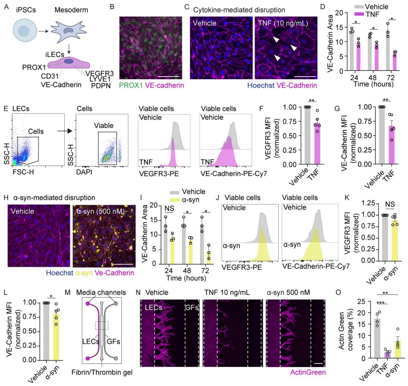
The third tier was in vitro experiments: human induced pluripotent stem cell-derived lymphatic endothelial cells (iLEC) were treated with recombinant TNF and α-syn PFF, respectively, to assess the tight junction protein VE-cadherin, the receptor VEGFR3, and three-dimensional sprouting capacity.
Core Findings: Deposition, Immune Activation, and Selective Drainage Impairment
At the human tissue level, both the cerebrospinal fluid and dura mater of patients with Parkinson's disease showed α-syn seeding activity, whereas control samples did not; discrete α-syn-positive inclusions were visible in the dura mater, mostly near Lyve1+ lymphatic vessels. No similar deposits were seen in Alzheimer's disease control tissue.
In the mouse acute experiments, injected α-syn accumulated mainly in the cerebrospinal fluid entry region around the dural sinuses, was taken up by monocytes, macrophages, and dendritic cells, and reached the deep cervical lymph nodes along meningeal lymphatic vessels. After 96 hours, dural macrophages increased, and the drainage of 3 kDa dextran and 45 kDa ovalbumin to the cervical lymph nodes decreased significantly, whereas 0.6 kDa cadaverine showed little change.
The chronic model reproduced a similar phenotype: macromolecular drainage declined at 1 month and persisted at 2 months; the connection pattern of meningeal lymphatic vessels shifted toward a "button-like" form. Notably, no α-syn deposition, gliosis, dopaminergic neuron loss, or behavioral abnormalities were observed in the brain parenchyma of these mice.
Innovation or Breakthrough: The Double Hit of TNF and α-syn
The study further pinpointed the mediator. Multiplex cytokine profiling of cerebrospinal fluid in the chronic model showed that TNF was significantly elevated, whereas IFNγ and IL-1β were unchanged. Reanalysis of published single-cell data also showed upregulated Tnf expression in meningeal and perivascular macrophages; in patient dura mater, α-syn-containing IBA1+ macrophages were positive for TNF immunoreactivity.
In vitro experiments provided the mechanism: after TNF treatment of iLEC, VE-cadherin junctions progressively disassembled, surface VE-cadherin and VEGFR3 decreased, and three-dimensional sprouting was impaired; α-syn PFF likewise disrupted VE-cadherin junctions and inhibited sprouting, but did not cause obvious NF-κB nuclear translocation, nor did it induce iLEC to release TNF themselves.
This means lymphatic dysfunction has two paths: macrophage-derived TNF indirectly damages lymphatic endothelium, whereas α-syn aggregates act directly on endothelial cells. The two partially overlap but are not entirely equivalent.
Practical Implications: Bringing Brain Borders into the Pathological Chain
This study recasts the dura mater from a "passive drainage interface" into an "interface actively participating in pathology." It suggests that clearance impairment in Parkinson's disease may appear before brain parenchymal pathology and be driven jointly by extracellular α-syn and local immunity.
For translational research, meningeal lymphatic drainage, border macrophage activation, and TNF signaling all become measurable dimensions for intervention. But the authors also explicitly noted limitations: human tissue sample sizes were limited, and the two mouse models cannot fully reproduce the entire process of α-syn from neuronal origin to spread; whether the basal meninges and nasopharyngeal lymphatic pathways are similarly affected remains to be verified.
Summary and Reflections
This study provides a mechanistic clue rather than a clinical conclusion: α-syn aggregates in the cerebrospinal fluid and dura mater are sufficient to cause immune activation and selective drainage impairment in animal models, and this impairment can exist independently of brain parenchymal pathology.
If clearance impairment can indeed occur before neuronal damage, could imaging or fluid biomarkers assessing meningeal lymphatic function become an earlier observation window? And given the persistent presence of α-syn, is blocking TNF alone sufficient to restore drainage? These questions deserve serious consideration in the design of the next round of experiments.