


The Alzheimer's disease (AD) risk gene CASS4 has long been considered associated with microglia, but its function in the brain remains unclear. A mouse study published in the Journal of Experimental Medicine shows that knocking out Cass4 aggravates Aβ deposition and neurite damage in the 5xFAD model and impairs cognitive flexibility; mechanistically, loss of CASS4 disrupts focal adhesion signaling in microglia, making it difficult for them to converge on Aβ plaques and compact them. The evidence comes from mice and in vitro cell experiments and is not yet a clinical conclusion.
Research background: a risk gene repeatedly hit yet rarely studied
Genome-wide association studies (GWAS) have repeatedly linked the CASS4 locus to late-onset AD risk. CASS4 encodes a scaffold protein belonging to the Cas family; it has no enzymatic activity itself and assembles protein complexes through multiple protein-binding domains.
The most widely known function of the Cas family is regulating focal adhesions. Focal adhesions are actin-rich cytoskeletal structures that give the cell membrane protrusions and traction, enabling cells to migrate, change shape, and engulf material.
In the brain, Cass4 is mainly expressed by microglia. Microglia are the brain's resident immune cells; they migrate to the surface of Aβ plaques to form a barrier and compact loose Aβ fibrils into dense plaques, thereby reducing the toxic surface area in contact with neurons.
However, previous research on CASS4 has mostly been confined to cell lines and oncology, and its role in AD pathology is almost a blank.
Research objective: does Cass4 deficiency alter the amyloidosis process
The authors generated whole-body Cass4 knockout mice and crossed them with the 5xFAD amyloidosis model to systematically assess Aβ burden, neurite health, cognitive behavior, and microglial responses.
The study further generated a microglia-specific conditional knockout model to determine whether the phenotype arises from the central nervous system's resident macrophages themselves.
Research methods (brief)
Morphologically, Thioflavin S was used to label dense-core plaques, 6E10 to label diffuse Aβ, and APP and ATG9 to label dystrophic neurites, with plaque volume, number, sphericity, and intensity quantified.
At the molecular level, single-nucleus RNA sequencing was performed on the hippocampus, and phosphorylated protein antibody array analysis was performed on magnetic bead-sorted microglia. At the functional level, the Morris water maze, in vitro scratch assay, primary microglial spreading assay, and in vivo two-photon laser injury imaging were used.
Core findings: more plaques, looser plaques, more neuronal damage
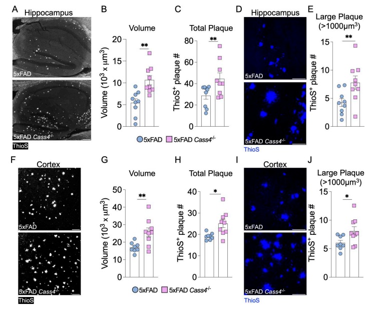
In the hippocampus and cortex of 5-month-old 5xFAD Cass4 knockout mice, the total volume and number of ThioS-positive plaques were increased, and there were also more large plaques with diameters corresponding to volumes exceeding 1000 μm³ (Figure 1).
Neurite damage was aggravated in parallel: APP-positive punctate structures around plaques increased, and ATG9 deposition was elevated in the hippocampus and cortex (Figure 2). Single-nucleus sequencing showed gene expression changes in both excitatory and inhibitory neurons.
Behaviorally, 4-month-old mice performed normally in the water maze place navigation and memory probe trials, but their ability to find a new location after platform relocation was markedly impaired, suggesting a selective deficit in cognitive flexibility (Figure 3).
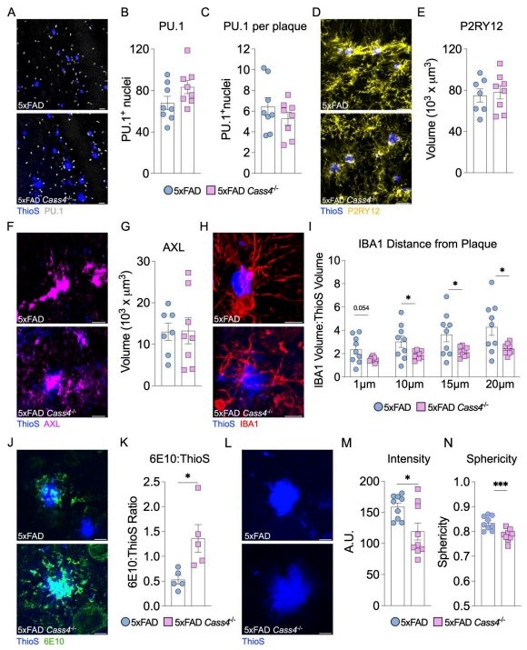
Microglial number, proliferation, DAM phenotype, and phagocytic capacity showed no obvious changes. What actually changed was "approach": IBA1 volume around plaques decreased, the 6E10-to-ThioS ratio increased, and plaque intensity and sphericity decreased (Figure 4).
Microglia-specific knockout reproduced the increased Aβ burden, poorer plaque compaction, and neurite damage, indicating that CASS4 acts in the central nervous system's resident macrophages.
Innovation or breakthrough: connecting CASS4 to the focal adhesion pathway
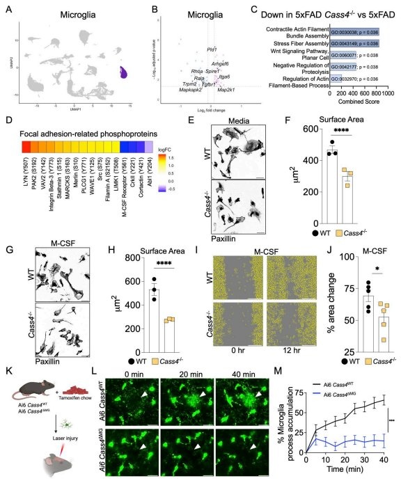
Phosphorylated protein array analysis showed that of 93 differentially phosphorylated proteins, approximately 17% were core molecules of focal adhesion signaling. Phosphorylation of CrkII, cortactin, and Abl1 decreased, while phosphorylation of integrin β3, Src, filamin A, and LYN increased; the latter may represent compensation.
Functional validation was consistent with the signaling changes: Cass4-deficient microglia showed reduced spreading area on laminin and a weakened response to M-CSF; macrophage scratch wound healing was slower; and after in vivo laser injury, the convergence of microglial processes toward the injury site was markedly blunted (Figure 6).
Practical significance: plaque "compaction" is also a protective mechanism
This study links AD genetic risk, microglial motility, and focal adhesion biology into a single thread. It suggests that, beyond plaque number, the tightness of plaques and the containment efficiency of microglia also affect surrounding neurons.
For translational research, the CASS4 pathway offers an interventional perspective different from Aβ clearance: improving microglial adhesion and migration may indirectly reduce neurotoxicity. However, the authors also note that how human AD-associated CASS4 single-nucleotide polymorphisms affect protein function remains to be studied, and this paper used a knockout model rather than a point-mutation model.
Summary and reflections
In the mouse model, loss of CASS4 does not change microglial number or "activation level"; instead, it makes them less able to approach and compact Aβ plaques, thereby aggravating neurite damage and cognitive flexibility deficits. This provides new clues for understanding how AD risk genes influence pathology through cell mechanics.
A question worth considering: if the core problem of microglia is not "insufficient numbers" but "inability to attach," then how should strategies targeting focal adhesion signaling enhance containment while avoiding excessive inflammation? This remains a question that requires experimental answers.