


Overactivation of dendritic cells is a key component of multiple autoimmune diseases, yet its metabolic regulatory mechanisms have long remained unclear. A study published in the Journal of Experimental Medicine (J. Exp. Med. 2026;223:e20252558) found that Vanin enzymes drive cholesterol and lipid raft accumulation through hydrolysis of pantethine, thereby promoting dendritic cell activation; in a psoriasis-like mouse model and patient-derived cells, either pantethine supplementation or Vanin inhibition attenuated this process. The evidence comes mainly from mouse models, in vitro cell experiments, and a small investigator-initiated clinical trial.
Research Background: Where Do Overactivated Dendritic Cells Come From?
Dendritic cells (DCs) are professional antigen-presenting cells responsible for bridging innate and adaptive immunity. Upon stimulation by antigens or danger signals, DCs upregulate CD80, CD86, and MHC molecules and secrete cytokines, thereby initiating T cell responses.
The problem is that once this program spirals out of control, it disrupts immune homeostasis. Abnormal DC activation has been observed in autoimmune diseases such as psoriasis and systemic lupus erythematosus. DCs are therefore regarded as attractive targets for intervention.
Recent studies suggest that metabolic pathways are regulators of DC activation, with glycolysis, amino acid metabolism, and fatty acid metabolism all participating. However, direct evidence is still lacking as to whether these metabolic nodes can be translated into therapeutic strategies. The Vanin enzyme family (VNN1, VNN2, VNN3) can hydrolyze pantethine to generate pantothenic acid and cysteamine; it has previously been discussed mostly as a marker associated with infection or tumors, and its role in DC function has been almost entirely unexplored.
Research Objective: Testing Whether a Metabolic Axis Regulates DCs
The core hypothesis proposed by the research team was that the Vanin–pantethine axis is a key metabolic regulatory pathway in DC activation, and that intervening in this axis may alleviate autoimmune inflammation.
Around this hypothesis, the research proceeded at three levels: first, seeking clues in patient transcriptomic data; then, verifying causality using gene knockout and pharmacological tools; and finally, testing translational potential in animal models and patient-derived cells.
Research Methods (Brief)
The study first analyzed public transcriptomic data of peripheral blood mononuclear cells and myeloid DCs from patients with multiple autoimmune diseases, comparing the expression of Vanin family genes.
At the cellular level, mouse bone marrow-derived DCs (BMDCs) and human monocyte-derived DCs were used; functional assessment was performed through siRNA knockdown, gene knockout, TLR agonist stimulation, combined with antigen presentation assays and OT-II T cell co-culture.
At the animal level, DC-specific Vnn3 knockout mice were generated, and phenotypes were observed in an imiquimod (IMQ)-induced psoriasis-like dermatitis model; interventions were carried out via both pantethine supplementation and the Vanin inhibitor RR6.
At the mechanistic level, metabolites were detected by liquid chromatography-mass spectrometry, cholesterol and lipid rafts were labeled with Filipin III and cholera toxin B subunit, and membrane localization of TLR4 as well as downstream NF-κB and MAPK signaling were tracked.
Core Findings: Less Pantethine, More Cholesterol Accumulation
First, Vanin genes are significantly upregulated in activated DCs. In peripheral blood and DC transcriptomes from patients with multiple autoimmune diseases, VNN1, VNN2, and VNN3 expression was elevated; in mouse BMDCs stimulated with LPS, Vnn3 was the predominantly upregulated isoform. In psoriatic lesions, VNN1 and VNN3 were positively correlated with activated DC signature genes.
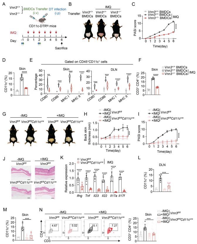
Second, Vnn3 deficiency impairs DC function. After knockout or silencing of Vnn3, DC antigen uptake and presentation decreased, CD80, CD86, and MHC molecule expression was reduced, and the ability to initiate T cell proliferation and secretion of IFN-γ, IL-17, and IL-4 was weakened. Notably, this suppression did not represent a shift toward a tolerogenic phenotype—regulatory T cell induction did not increase, nor were IL-10 and PD-L1 elevated.
Third, animal models verified causality. Mice with DC-specific Vnn3 deficiency exhibited milder lesions, lower PASI scores, and reduced epidermal hyperplasia and inflammatory infiltration under IMQ induction, with decreased expression of Ifng, Tnf, Il23, Il22, Il17a, and Il17f in the skin.
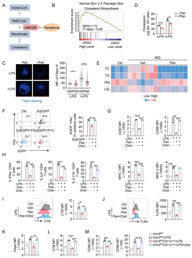
Fourth, the mechanism centers on cholesterol and lipid rafts. Vanin hydrolyzes pantethine, and pantethine can inhibit HMG-CoA reductase. Vnn3 deficiency led to accumulation of pantethine in cells and plasma, and decreased cholesterol and lipid raft content in DCs; supplementation with exogenous cholesterol reversed the inhibition of DC activation by pantethine. After lipid raft integrity was impaired, enrichment of TLR4 on the membrane decreased, and downstream NF-κB and MAPK signaling was attenuated.
Fifth, pharmacological inhibition is similarly effective. The Vanin inhibitor RR6 elevated intracellular pantethine, reduced cholesterol and lipid rafts, weakened DC presentation and T cell activation capacity, and alleviated dermatitis in the IMQ model.
Innovations or Breakthroughs
The novelty of this work lies in connecting a metabolic pathway to a specific cellular mechanism: Vanin is not a vague inflammatory marker, but rather directly influences TLR4 signal transduction by consuming pantethine, raising membrane cholesterol, and remodeling lipid rafts.
Another noteworthy observation is that Vnn3 deficiency did not push DCs toward a tolerogenic phenotype, but rather weakened their activation capacity overall. This has implications for understanding the distinction between "suppressing DCs" and "inducing tolerance."
In addition, the study verified the inhibitory effects of pantethine and RR6 in patient-derived psoriatic and SLE cells, and reported a small clinical trial: adding oral pantethine to a topical regimen resulted in greater reductions in PASI and PGA scores at 12 weeks, with more pronounced benefit in patients with comorbid dyslipidemia, and no treatment-related adverse reactions were observed. The authors also emphasized that this trial was a feasibility validation and cannot directly prove that the intrinsic DC pathway is the sole reason for clinical benefit.
Practical Implications: Another Approach to Metabolic Intervention
For clinical and translational research, this study suggests that cholesterol metabolism may be an actionable immunomodulatory node. Pantethine itself is a low-molecular-weight thiol compound long used for lipid management, with a relatively long safety record, which facilitates exploration of "old molecules, new uses."
However, interpretation requires restraint. The mechanistic evidence comes mainly from mouse BMDCs and the IMQ model, and the IMQ model cannot fully represent human psoriasis; the patient cell experiments are in vitro results; and the clinical trial had a small sample size, was single-center, and had short-term follow-up. Vanin is expressed in multiple immune and stromal cells, and whether its effects are confined to DCs was explicitly left by the authors for future research.
Summary and Reflections
This study places the Vanin–pantethine–cholesterol axis at the center of DC activation, providing a mechanistically supported candidate direction for metabolic intervention in autoimmune diseases. From transcriptomic clues to gene knockout, pharmacological intervention, patient cells, and finally small-scale clinical observations, the evidence chain is relatively complete, but the applicable boundaries of each link require clarification by future research.
One question worth considering: if cholesterol lowering itself can modulate DC function, might statins, which are widely used clinically, also produce similar immunomodulatory effects in specific DC subsets and inflammatory contexts? The results reported in existing studies are inconsistent, and this may be precisely where controlled experiments are needed next to provide answers.