



The combination of suprachoroidal injection and RPE-specific promoters is rewriting the rules of retinal gene delivery.
Dry age-related macular degeneration (dAMD) is a leading cause of irreversible vision loss in the elderly. At the core of its pathology is the long-term assault of retinal pigment epithelium (RPE) cells by oxidative stress and chronic inflammation. However, how to safely and precisely deliver therapeutic genes to the RPE has long been a technical bottleneck in the field.
A study published in Journal of Controlled Release in 2026 proposed a "dual-restriction" strategy: achieving anatomical targeting through suprachoroidal administration, and then achieving transcriptional-level restriction via the RPE-specific Best1 promoter. This combination provides a new design paradigm for gene therapy of dAMD.
I. Starting from Mechanism: Recovering the Lost "Molecular Brake"
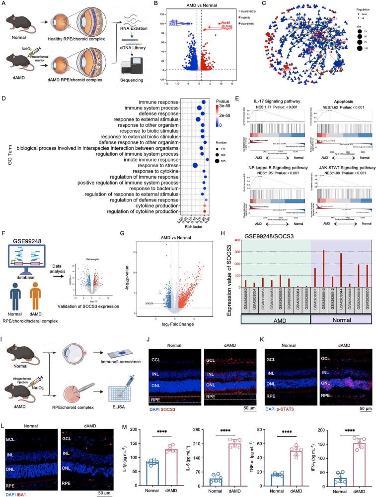
The research team first performed transcriptomic sequencing on a sodium iodate-induced dAMD mouse model. In the RPE/choroid complex, a total of 1515 differentially expressed genes were identified, among which SOCS3 was significantly downregulated. SOCS3 is a key negative feedback regulator of the JAK-STAT signaling pathway, and can be described as the "molecular brake" of the inflammatory response.
This finding was validated in human samples. Analysis of the public dataset GSE99248 showed that in the RPE/choroid/sclera complex of AMD patients, SOCS3 expression was likewise significantly reduced. At the same time, p-STAT3 levels were elevated, suggesting abnormal activation of the JAK-STAT pathway.
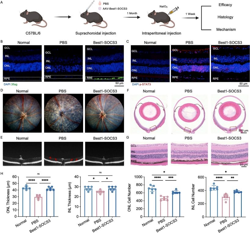
The mechanistic chain gradually became clear: loss of SOCS3 leads to sustained phosphorylation of STAT3, which in turn amplifies the inflammatory response, disrupts mitochondrial function, and exacerbates oxidative stress, ultimately driving RPE degeneration. Restoring SOCS3 is equivalent to reinstalling this brake system.
II. The Battle Over Delivery Routes: Why Is Suprachoroidal Superior?
The RPE is located in the outermost layer of the retina, and traditional administration methods each have their shortcomings. Intravitreal injection is simple to perform, but is limited by the inner limiting membrane barrier, making it difficult to effectively transduce the outer retina and RPE. Although subretinal injection can directly contact the RPE, it requires penetrating the retina and creating a temporary retinal detachment, carrying risks of hemorrhage and local damage.
The study compared three administration routes. The results showed that after intravitreal injection, transgene expression was mainly confined to the ganglion cell layer and inner nuclear layer, with almost no signal in the RPE.
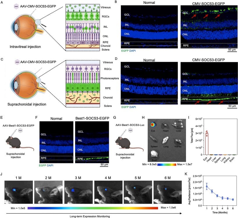
Suprachoroidal injection is completely different. AAV enters through the potential space between the sclera and choroid, and without needing to penetrate the retina, can accumulate at the choroid-RPE interface. Under the drive of the CMV promoter, suprachoroidal administration was already able to achieve predominantly RPE transgene expression, although a small amount of off-target signal remained in the adjacent retinal layers.
This suggests that anatomical targeting alone is not enough; a second layer of restriction is needed.
III. The Best1 Promoter: Locking Expression into the RPE
The Best1 gene encodes the bestrophin-1 protein, which is specifically and highly expressed in RPE cells. The research team constructed an AAV vector driven by the Best1 promoter and conducted a head-to-head comparison with the broad-spectrum CMV promoter.
At the same dose and administration route, the CMV promoter produced relatively widespread outer retina/RPE expression, with signal able to spread beyond the RPE. In contrast, the Best1 promoter strictly confined expression to the RPE layer, with significantly reduced off-target signal in the neural retina. TUNEL staining further showed that retinal cell apoptosis in the Best1 group was markedly less than in the CMV group.
This design has deeper considerations. STAT3 signaling has context-dependent functions in different retinal cells: under pathological conditions it promotes inflammation, but in neurons it may support stress responses and neuroprotection. If SOCS3 were broadly expressed in the neural retina, it might inadvertently harm these protective pathways.
IV. Long-Term Expression and Safety: Key Data for Clinical Translation
Beyond efficacy, the researchers also systematically evaluated durability and safety. Luciferase reporter genes showed that after a single suprachoroidal injection, local ocular expression could persist for at least 6 months. Biodistribution analysis indicated that the vector was mainly confined to the injected eye, with extremely low signal in the heart, liver, spleen, lung, kidney, and brain.
The safety assessment covered intraocular pressure, electroretinography, visual cliff behavioral testing, blood counts, liver and kidney function, and histology of major organs. At the working dose of 2×10¹⁰ vg/eye, no obvious toxicity was found. Notably, neutralizing antibodies against the AAV capsid were detectable in serum, but no ocular inflammatory cell infiltration or systemic cytokine elevation was observed.
Dose-exploration experiments also revealed an interesting plateau phenomenon: RPE transgene expression continued to rise from 5×10⁹ to 2×10¹⁰ vg/eye, but further increasing the dose to 4×10¹⁰ vg/eye produced no additional gain. This suggests that 2×10¹⁰ vg/eye is the minimum effective dose approaching maximal expression.
Conclusion: From "Can It Be Delivered" to "How Precisely Can It Be Delivered"
The value of this study lies in upgrading the question of "can it be delivered to the RPE" to "how to achieve dual precision control at both the anatomical and transcriptional levels." Suprachoroidal administration addresses the physical barrier, while the Best1 promoter addresses cell specificity; the combination of the two significantly improves the therapeutic window.
But many questions remain unresolved. Can SOCS3 still be effective in late-stage geographic atrophy, in the face of massive RPE death? Will Best1 promoter activity decline after chronic oxidative stress causes RPE dedifferentiation? How should the impact of neutralizing antibodies on repeated dosing be managed?
When we design the next generation of AAV processes, should we also shift from the single-dimensional thinking of "vector delivery efficiency" to multi-dimensional optimization of "spatial distribution + transcriptional regulation + temporal durability"? This may be the real proposition this paper leaves for process developers.