



A 3-year-old child with infantile Krabbe disease developed a GLI1-altered tumor paravertebral to L2–L3 after receiving AAV gene therapy. Long-read sequencing showed that a subgenomic vector fragment lacking the GALC transgene and poly(A) signal integrated into GLI1 intron 1, driving full-length GLI1 expression via splicing. This case highlights the significance of vector purity and cassette design for long-term safety.
Case Background: Gene Therapy After Two Transplants
Krabbe disease is a rare autosomal recessive disorder. Mutations in the GALC gene cause galactocerebrosidase deficiency, accumulation of the lipid psychosine, and widespread demyelination and neurodegeneration of the central and peripheral nervous systems. AAV-mediated gene therapy is therefore regarded as a promising therapeutic strategy.
The child carried biallelic GALC variants (p.Arg220Ter and p.Tyr319Cys). He underwent two umbilical cord blood transplants at 5 months and 9 months of age, each preceded by myeloablative alkylator conditioning.
On day 29 after the second transplant, he received intravenous FBX-101 at a dose of 3×10¹³ vector genomes/kg body weight, as part of the RESKUE trial. FBX-101 is an AAVrh.10 capsid vector flanked by AAV2 inverted terminal repeats (ITRs), carrying GALC under the control of a CAG promoter—composed of the cytomegalovirus enhancer and the chicken β-actin promoter.
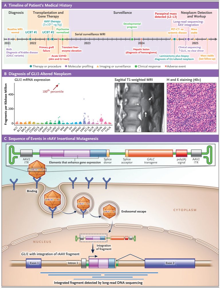
After treatment, the patient's immune reconstitution remained poor; he received donor lymphocyte infusions and immunosuppressive therapy for graft-versus-host disease. Twenty-six months later, surveillance MRI incidentally detected a paravertebral mass at L2–L3.
Molecular Features of the Tumor: GLI1 Activation
Biopsy revealed a small round blue cell tumor with focal epithelioid, nested appearance. The specimen was positive for CD56 and S100, in situ hybridization showed strong GLI1 positivity, and the Ki-67 index was below 10%. The diagnosis pointed to a GLI1-altered neoplasm.
GLI1 mRNA levels ranked at the 100th percentile among 5,143 samples in the institution's pan-cancer cohort, accompanied by PTCH1 overexpression. Targeted sequencing showed a stable diploid genome, with no oncogenic mutations or fusions detected.
This meant that the tumor was not driven by classic point mutations or gene fusions, but by another mechanism.
Long-Read Sequencing Pinpoints the Integration Event
Nanopore long-read sequencing identified a clonal 1038 bp insertion in GLI1 intron 1, present in 44% of reads, flanked by a 115 bp target site duplication.
The integrated fragment was a subgenomic vector fragment containing a partial AAV2 ITR, the cytomegalovirus enhancer, the chicken β-actin promoter, exon 1, and a splice donor; it lacked the GALC transgene and the poly(A) signal.
This splice donor generated a chimeric transcript linking chicken β-actin exon 1 to GLI1, driving full-length GLI1 expression. Allele-specific hypomethylation confined to the integrated allele further established the integration as the direct cause of GLI1 activation.
The authors noted that AAV vectors are predominantly non-integrating and have an established safety record in thousands of recipients. However, this case resembles a previously reported case by Ahrens-Nicklas et al.: the integrants were truncated, rearranged subgenomic structures that retained the CAG promoter-enhancer and a splice donor, activating a developmental transcription factor.
Manufacturing Perspective: A Dual Warning About Impurities and Cassette Design
A key detail is that the integrant in this case contained neither the transgene nor the poly(A) sequence. The authors proposed that "snapback" or promoter-only vector species generated during manufacturing may be the integration substrate.
This points to two manufacturing-level issues. First, vector purity—truncated, transgene-free subgenomic species generated during packaging may serve as potential integration substrates. Second, cassette design—elements containing a splice donor, once integrated near a host gene, may generate chimeric transcripts.
The authors also discussed the potential contribution of the transplant context in this case: DNA-damaging therapy and immunosuppression may have created an environment conducive to clonal expansion. The integration event was promptly reported to the treating physician and the U.S. Food and Drug Administration. At last follow-up, the patient remained asymptomatic.
Implications for AAV Manufacturing
The core recommendation of this letter is clear: splice-donor-free cassette designs are needed, along with attention to vector purity and extended genomic monitoring after AAV gene therapy.
For process development, it elevates "vector purity" from a release specification to a safety issue. Subgenomic impurities, truncated genomes, promoter-only species—components that may be tolerated in routine quality control—take on new weight in the context of integration-driven tumorigenesis.
At the same time, seemingly harmless splice donor elements in cassette design may become a switch that activates adjacent proto-oncogenes in an integration event. Sequence choices at the design stage are directly relevant to long-term safety.
One question worth persistently pursuing is: among predominantly non-integrating AAV vectors, which class of subgenomic impurity, at what dose and in what host context, is capable of integrating and driving clonal expansion? Answering this may require more systematic vector component characterization and long-term follow-up data.