



Adeno-associated virus (AAV) vectors are widely used in gene therapy, but their natural tropism is too broad. This creates a long-standing contradiction: to make them infect only a certain cell type, one often has to modify the capsid; yet once the capsid is altered, yield, assembly, and infectivity are often affected as well.
A recent study on neuroblastoma offers a different approach. The researchers introduced the SpyTag/SpyCatcher covalent conjugation system into the AAV9 capsid to redirect viral tropism using antibody fragments. The process details involved are of considerable reference value to vector developers.
The design logic of a "neutral" capsid
The study did not choose ordinary AAV9, but rather the AAV9-W503A mutant. This mutation disrupts the binding between the capsid and galactose, causing the virus to almost completely lose its natural infectivity.
In other words, this is a "blank packaging vector." Its natural tropism has been emptied out, and whom it subsequently infects is entirely determined by the targeting molecule attached to its surface.
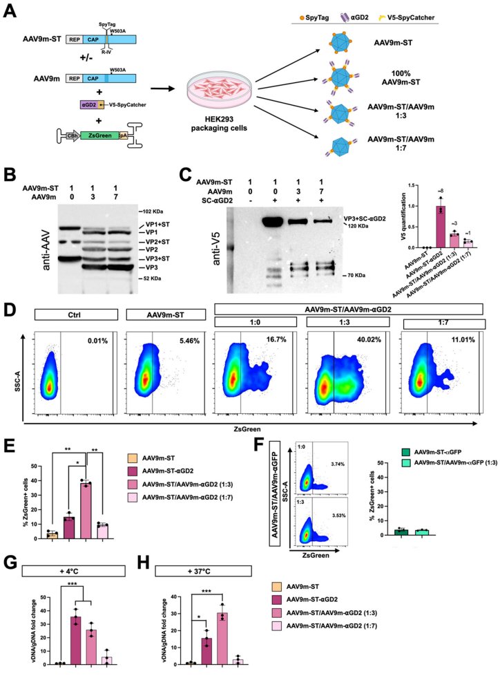
The researchers inserted the mere 13-amino-acid SpyTag at position 453 of capsid variable region IV. This site had previously been shown to be editable without affecting viral yield or infectivity. The corresponding SpyCatcher was fused to an anti-GD2 scFv.
When the two meet, they spontaneously form a covalent isopeptide bond. The entire process requires no additional chemical crosslinking.
Two conjugation routes, two outcomes
The study compared two process routes.
The first was "post-production conjugation": viral particles and purified SpyCatcher-scFv protein were prepared separately, then mixed and incubated at different ratios.
The results were not ideal. The scFv fusion protein recovered from the supernatant showed a molecular weight shift, which was confirmed by deglycosylation experiments to be caused by glycosylation. More critically, the conjugation efficiency was low—even at an extreme ratio of 1:800, a large amount of unconjugated protein remained, and it was difficult to remove even by filtration purification.
The second was "co-expression conjugation": the plasmid encoding scFv-SpyCatcher was co-transfected into HEK293T cells along with the AAV packaging plasmids.
This route completes the covalent linkage intracellularly, and after iodixanol gradient purification there is almost no free protein. The intracellularly expressed scFv does not undergo glycosylation, and the protein purification step is also eliminated. Multiple independent preparations showed stable viral titers.
Based on these results, the researchers subsequently adopted the co-expression route for all experiments.
More conjugation, worse infection
A counterintuitive finding emerged regarding conjugation density.
Western blot quantification showed that under non-chimeric conditions, approximately 13±3% of VP3 was conjugated; calculated at approximately 50 VP3 per capsid, each capsid on average carried 7–8 scFv.
But high-density conjugation instead inhibited infection. On GD2-positive SH-SY5Y cells, the transduction efficiency of the non-chimeric vector was less than 10%.
The researchers speculated that this was due to steric hindrance—overly dense surface molecules shielding the capsid's recognition of co-receptors. Binding experiments at 4°C showed that the membrane binding of chimeric capsids actually decreased, indicating that the problem was not in adsorption, but in the subsequent entry or intracellular trafficking steps.
1:3 chimeric: an empirical balance point
To verify the steric hindrance hypothesis, the researchers mixed Rep-Cap plasmids with and without SpyTag at different ratios to prepare chimeric capsids.
At a 1:3 ratio, the number of scFv per capsid dropped to 2–3, yet the transduction efficiency increased more than 6-fold compared with non-chimeric conditions. At a 1:7 ratio, the conjugation number was less than 1, and the efficiency fell back.
This 1:3 configuration was named AAV-STITCHαGD2. Its specificity was also verified: when switched to an irrelevant anti-GFP scFv, transduction completely disappeared; in HEK293T cells that originally did not express GD2, after forced expression of GD2 synthase, only GD2-positive cells were transduced.
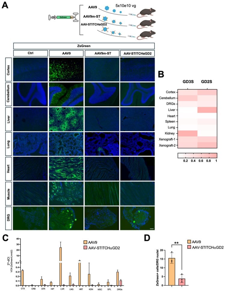
In vivo distribution further confirmed the specificity. After intravenous injection, no ZsGreen expression was detectable in liver, heart, muscle, brain, or lung, with the sole exception of the dorsal root ganglion—consistent with the known GD2 expression at that site.
Process implications and unresolved questions
The value of this work lies not only in the therapeutic results, but even more in the process principles it reveals.
First, there is an optimal range for conjugation density. Surface modification is not a case of more being better; excess ligand can interfere with the capsid's natural entry pathway. 1:3 is a robust starting point, but the researchers explicitly stated that it may not be universally applicable.
Second, the co-expression route moves conjugation from "post-production" to "during production," simplifying the workflow and also avoiding the heterogeneity brought about by glycosylation of secreted proteins. This is advantageous for scale-up manufacturing.
Third, using an infection-defective capsid as the chassis can keep off-target risk at a relatively low level, at the cost of necessarily relying on exogenous targeting molecules to take effect.
There are also many questions still to be answered: at which step of the transduction cycle does excess scFv actually block? Can the chimeric ratio be rationally predicted rather than relying on empirical screening? When the targeting molecule is switched from scFv to another ligand, does this ratio still hold?
For those working on vector process development, these questions may be more worth pondering than the efficacy data.